


| Issue |
Vis Cancer Med
Volume 7, 2026
|
|
|---|---|---|
| Article Number | 3 | |
| Number of page(s) | 12 | |
| DOI | https://doi.org/10.1051/vcm/2026002 | |
| Published online | 09 June 2026 | |
Review Article
Multifunctional nanoparticles in colorectal cancer therapy: Advances in targeted drug delivery systems
Department of Chemistry and Biochemistry, School of Sciences, JAIN (Deemed-to-be University), JC Road, 34, 1st Cross Rd, Bengaluru, 560027, Karnataka, India
* Corresponding author: This email address is being protected from spambots. You need JavaScript enabled to view it.
Received:
25
July
2025
Accepted:
26
January
2026
Abstract
Colorectal cancer (CRC) is a major global health concern with a high fatality rate, particularly in its late stages. Traditional treatments, such as surgery and chemotherapy, have been reported as unsuccessful in some cases due to adverse side effects, lack of pharmaceutical specificity, and drug resistance. Nanoparticle-mediated delivery of medications has been presented as a unique method for increasing treatment efficacy while reducing systemic toxicity. Nanoparticles (NPs), which comprise liposomes, polymers, dendrimers, silica nanoparticles, and nanoemulsions, provide controlled drug release, improved bioavailability, and tumour-specific therapeutic targeting. Their physicochemical properties enable them to be functionalised with greater specificity while reducing off-target effects. This review combines multiple nanoparticle-based drug delivery techniques for CRC therapy, assessing their merits, shortcomings, and potential future applications. Though nanomedicine has shown great promise in preclinical and clinical trials, there are obstacles to optimising targeted approaches and identifying specific molecular biomarkers. Continued research into nanobioconjugation and multifunctional nanocarriers can pave the road for more effective and personalised CRC treatment.
Key words: Colorectal cancer (CRC) / nanoparticles / Drug delivery / Nanobioconjugation
© The Authors, published by EDP Sciences, 2026
 This is an Open Access article distributed under the terms of the Creative Commons Attribution License (https://creativecommons.org/licenses/by/4.0), which permits unrestricted use, distribution, and reproduction in any medium, provided the original work is properly cited.
Introduction
Colorectal cancer (CRC) ranks among the top causes of cancer-related mortality globally in both men and women. [1]. CRC is a major global health concern, ranking as the third most common cancer worldwide, with over 1.9 million cases reported in 2022 (1,045,413 in males and 826,706 in females). In India, CRC is the fourth most common cancer among both sexes, with 64,863 new cases and 38,367 deaths reported in 2022 [2]. In the United States, it is the third most diagnosed cancer and the third leading cause of cancer-related deaths in both men and women. According to the American Cancer Society (ACS), an estimated 154,000 new CRC cases are expected in the U.S. in 2025. Globally, the lifetime risk of developing CRC is approximately 1 in 24 for men and 1 in 26 for women [3]. The prognosis of colorectal cancer largely depends on the stage at which it is diagnosed. Early diagnosis, including colonoscopies, creates more possibilities of survival [4]. However, mortality rates are higher in less developed countries, predominantly in Asia, Africa, Latin America and the Caribbean etc., due to limited resources and inadequate healthcare infrastructure. Colorectal cancer rates are declining in high-income countries due to effective screening programmes. Early diagnosis and timely treatment significantly improve survival and quality of life (World Health Organization (WHO), 2023) [5]. Age constitutes one of the most important risk factors in the development of CRC, with adolescents (20–39 years according to ACS) and older age groups being the vulnerable populations [6]. In India, the age-standardized rate (ASR) is relatively low, at 7.2 per 100,000 males and 5.1 per 100,000 females. Indian cancer registries have seen an increasing trend with annual percent changes ranging between 20 and 124% [7]. However, the five-year survival rate for CRC patients in India is less than 40%, ranking low globally [8].
Nanoparticles have emerged as an important field of study in targeted medication delivery due to their capacity to deliver pharmaceuticals to specific places with sustained release while minimising negative effects in non-targeted areas [9]. NPs are characterized by at least one dimension under 100 nm and hold significant promise for drug delivery and medical treatments, especially in cancer therapy [10, 11].
Nanoparticle-based drug administration provides several advantages, including a longer circulation half-life, enhanced pharmacokinetics, the potential to combine high-dose medications, fewer adverse effects, and precise drug targeting to specific regions of the body [4]. Nanoparticles have attracted attention due to their biocompatibility, high surface-to-volume ratio, and biodegradability in biological contexts. Their different physicochemical properties enable conjugation and functionalisation for targeted drug delivery. Metals, metal oxides, polymers, and biopolymers are nanoscale materials with improved nanodrug formulations for targeted therapy. Nanobioconjugation is essential in targeted cancer therapy because it covalently binds cancer receptors, antibodies, ligands, and cytotoxic medicines [12]. This improves drug specificity, reduces side effects, and increases therapeutic efficacy [13]. Conventional therapies for CRC, including chemotherapy, radiotherapy and surgery, prove to be insufficient for successful treatment due to systemic toxicity, drug non-specificity and drug resistance. Such restrictions may have adverse effects and result in inadequate therapeutic effects. This review focuses on different types of advanced nanoparticle-based drug delivery systems as a promising alternative. By enabling targeted delivery, improved bioavailability, and minimized off-target effects, these systems aim to enhance the efficacy and safety of colorectal cancer therapy.
Pathophysiology
Colorectal cancer (CRC) develops through a multistep process in which normal colonic epithelium gradually acquires genetic and epigenetic alterations, leading first to precancerous adenomatous polyps and ultimately to invasive carcinoma and becoming malignant by spreading to distant organs, such as the lungs, liver, and bone marrow, as shown in Figure 1 [14]. This progression is driven by the accumulation of mutations, either acquired somatically or inherited, that disrupt critical cellular pathways, resulting in uncontrolled cell growth, evasion of apoptosis, angiogenesis and enhanced metastatic potential [15]. Clinical evidence shows that colorectal cancer frequently arises from adenomatous polyps, which can persist and accumulate abnormal cellular changes over a decade or more before becoming malignant [16]. CRC involves dynamic interaction among various molecular pathways, majorly chromosomal instability (CIN), characterized by aneuploidy and mutations in genes such as adenomatous polyposis coli (APC), microsatellite instability (MSI) due to defective DNA mismatch repair, and the CpG island methylator phenotype (CIMP), which alters gene expression through DNA methylation. In sporadic CRC, these changes typically begin with mutations in APC and KRAS, whereas in colitis-associated CRC, chronic inflammation plays a central role by inducing cytokine release and field changes in the colonic mucosa, with earlier acquisition of p53 mutations and later involvement of APC/KRAS [17].
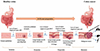 |
Figure 1 Development of colorectal cancer: Initiation, promotion, progression, local progression, metastasis and common metastatic sites [14]. |
Colitis-associated CRC is characterized by multifocal dysplasia and a unique mutational profile enriched in genes related to cell communication and adhesion, all of which may be linked with the dysregulated cytokines and inflammatory mediators associated with inflammatory bowel disease (IBD) [18]. Overall, these diverse molecular mechanisms not only underscore the heterogeneity of CRC but also highlight the importance of early polyp detection, personalized surveillance, and targeted therapeutic strategies based on underlying genetic and inflammatory profiles.
Diagnosis
Diagnosis of CRC is performed with a wide range of methods with varying sensitivity and specificity. Endoscopic methods, especially colonoscopy and sigmoidoscopy, afford the most efficient and also valuable diagnostic tools, obtaining direct visualization of tumours, as well as making it possible to take tissue biopsies and analyze them histologically. Although diagnostic, these methods are invasive and potentially may cause discomfort or complications of incidents such as haemorrhage and perforation. Other imaging methods, including computed tomography (CT), magnetic resonance imaging (MRI), and positron emission tomography combined with CT (PET/CT), are also utilized. Among these, PET/CT provides enhanced prognostic information, particularly valuable for evaluating treatment response [19]. Among non-invasive methods, faecal occult blood testing (FOBT) remains a widely used initial screening tool, and its sensitivity can be enhanced through repeated testing or the use of immunochemical-based methods like the faecal immunochemical test (FIT) [20]. Blood-based tumour markers, including Carcinoembryonic Antigen (CEA), Carbohydrate Antigen 19-9 (CA 19-9), Tissue Polypeptide-Specific Antigen (TPS) and Tumour-Associated Glycoprotein 72 (TAG-72), are also utilized; however, their limited sensitivity and specificity have prompted ongoing research into novel biomarkers such as lysosomal exoglycosidases [21]. Although a per rectum examination represents the simplest initial clinical approach, its diagnostic yield is considerably lower compared to endoscopic evaluations. To overcome the limitations of individual tests, combined screening strategies are recommended to enhance overall detection rates [22].
Treatment
Treatment modalities for colorectal cancer typically integrate surgery, cryosurgery, chemotherapy, immune-based therapies, radiation therapy, and targeted therapies [23]. The most prominent strategy in the therapeutic approach of chemotherapy is using one or a combination of various drugs to halt cancer cell division. However, in traditional chemotherapy, drugs reach places other than their targets and produce very harsh side effects like neutropenia, anaemia, hand-foot syndrome, diarrhoea, gastrointestinal toxicity, mucositis, nausea, vomiting, fatigue, haematologic disorders, and liver toxicity [24]. Recent advances in cancer treatment include strategies such as RNA-based therapies, oncolytic viral therapies, and approaches guided by specific biomarkers, all of which show considerable therapeutic promise [25]. However, even with these innovations, clinical outcomes remain suboptimal, largely due to persistent challenges like drug resistance, disease recurrence, metastasis, and significant adverse effects [26].
In CRC, immune checkpoint inhibition therapy has shown effectiveness, particularly in patients with tumours exhibiting high microsatellite instability (MSI-H) [27]. Effective CRC treatment not only requires controlling tumour cell growth but also involves pleiotropic mechanisms in the tumour microenvironment (TME) (Figure 2 and 3) [17, 28–30] to enhance anti-tumour immunity, thereby improving therapeutic outcomes.
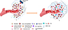 |
Figure 2 Schematic representation of the tumour microenvironment and its cellular composition of colorectal cancer [17, 28]. |
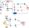 |
Figure 3 Pleiotropic mechanisms in the tumour microenvironment (TME). (a) Drug loaded NPs can inhibit the hypoxic tumor vasculature of colorectal cancer by targeting and reprogramming the multiple mechanisms: i) Myeloid-Derived Suppressor Cells (MDSCs) and T-regulatory cells (Tregs) inhibit cytotoxic T-lymphocytes (CTL) and drive tumour growth. ii) The conversion of M1 macrophages to M2 macrophages also leads to tumor growth. iii) TGFβ-induced fibroblasts activate Cancer-Associated Fibroblasts (CAFs), which promote tumor progression. iv) VEGF also stimulates abnormal tumour vasculature and cancer development. Drug loaded NPs can target all of these pathways to suppress tumour growth. NPs can reprogram M2 macrophages into M1 phenotypes, which reactivate the immune response and inhibit tumour growth. Drug loaded NPs can also activate mature DCs, leading to the activation of B cells through T cells and the production of antibodies that can target and eradicate tumour cells. These NP-stimulated pathways can thus overcome the inhibitory effects exerted by MDSCs on mature dendritic cells (DCs) and the inhibitory effects of Tregs on INF-γ, thus killing tumour cells. Additionally, NPs loaded with oxygen can inhibit the hypoxic tumour vasculature by inducing oxygen, thus reversing hypoxia and limiting tumour growth [29]. (b) Schematic representation of how drug-loaded nanoparticles modulate tumor-associated macrophages (TAMs) under hypoxic conditions. NPs inhibit TAM2 and promote TAM1 polarization, leading to increased IL-12 and decreased IL-10 production, which suppresses tumour vasculature and enhances anti-tumor immunity. Drug-loaded NPs can inhibit the conversion of TAM1 to TAM2 and reduce tumour growth. SiRNA-loaded NPs can also suppress TAM2 activity [30]. |
Colorectal cancer poses multiple physiological barriers, like vascular endothelial pores, heterogeneous blood supply, and an intricate architecture. The success of cancer therapy majorly depends upon the drug delivery method. Traditionally, several anticancer agents have been developed, but often these agents were unable to target the cancer cells specifically and gave rise to widespread toxicity and various side effects in the body. These challenges have become the point of focus for extensive research conducted to identify molecular biomarkers with both diagnostic and therapeutic potential. In addition, processes based on genetics and epigenetics play intertwined roles in driving cancer progression in colorectal cancer.
Circulation of nanoparticles in tumour microvessels
Exploring the mechanism of nanoparticle drug delivery from the vessel to the tumour through vascular extravasation is very important because this is a critical stage [31]. In general, Vascular extravasation of NPs in the tumour region can be mediated by the passive EPR effect. The Enhanced permeability arises from the large endothelial gaps in tumour vessels that allow the passage of NPs, and the enhanced retention results from a collapse of the lymphatic drainage system where nanocarriers (NCs) cannot be transported away [32].
Nanoparticles can easily pass through tumour microvessels rather than normal healthy vessels because of the following reasons: i) overexpression of VEGF on tumour cells and ii) abnormal basement membrane lining in tumour microvessels.
- i)
Overexpression of VEGF on tumour cells: Vascular endothelial growth factor (VEGF) drives the formation of abnormal, highly permeable tumour vasculature. By stimulating angiogenesis, VEGF generates microvessels with enlarged endothelial gaps (100–400 nm) and defective basement membranes, which markedly increase the permeability of the tumour’s blood supply. These structural changes amplify the Enhanced permeability and retention (EPR) effect, allowing nanoparticles typically 10–200 nm in diameter to extravasate from the tumour microvessels to the tumour interstitium. In contrast, normal vessels maintain low VEGF signalling, tight endothelial junctions and a continuous basement membrane, which together act as a stringent barrier that blocks NP extravasation. In addition to creating leaky vessels, VEGF-induced endothelial cells to overexpress VEGF receptors (e.g., VEGFR-2), as shown in Figure 4a. Nanoparticles can be functionalized with ligands or antibodies that bind these receptors, providing an active-targeting layer that further concentrates the carriers at the tumour vasculature and promotes receptor-mediated endocytosis. Such dual exploitation of VEGF-mediated vascular permeability and receptor targeting enhances both the amount of nanoparticle that enters the tumour and its retention within the malignant tissue [33].
- ii)
Abnormal basement membrane lining in tumour microvessels: The basement membrane surrounding tumour vessels is also markedly different from that of normal vasculature. In tumours it is often thin, heterogeneous, or partially degraded, allowing NPs that have crossed the endothelial gap to become trapped only transiently before further transvascular transport. Normal vessels have a uniform basement membrane in healthy tissue that sequesters NPs in the subendothelial space and strongly hinders their passage by forming a perivascular NPs pool, as shown in Figure 4b. The basement membrane is heterogeneous across different tumour regions, nanoparticles that can respond to the local microenvironment (e.g., pH-sensitive release) are better able to navigate these variable pathways. The combined effect of a compromised basement membrane and the leaky endothelial gaps generated by VEGF-driven angiogenesis amplifies the enhanced permeability and retention (EPR) effect, boosting nanoparticle accumulation within the tumour. Additionally, nanoparticles combined with collagen hydrolases are capable of degrading the basement membrane to release the trapped NPs, as shown in Figure 4c, and this may also lead to irreversible destruction of the basement membrane and increase the risk of cancer metastasis [34].
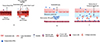 |
Figure 4 Normal microvessels with thick endothelial cells and tumour microvessels with leaky endothelial gaps due to overexpression of VEGFR (a) [33]. NPs are trapped in the subendothelial void by basement membrane surrounding vessels, forming a perivascular NPs pool that hinders their extravasation (b). Collagen hydrolase-mediated basement membrane destruction is used to increase NP extravasation (c) [34]. |
Nanoparticles in drug delivery systems for colorectal cancer
Nanoparticles were first introduced as drug delivery vehicles in the mid-1980s, when it was observed that they could preferentially accumulate within tumour tissues through a process now recognized as the Enhanced Permeability and Retention (EPR) effect [35]. This passive targeting mechanism marked a significant advancement in the use of nanoparticles for cancer treatment [36]. Due to their ability to selectively target tumours, achieve high levels of accumulation at disease sites, and maintain prolonged circulation within the bloodstream, nanoparticles have become promising tools in the development of cancer therapies [37]. Some of the nanocarriers used in drug delivery systems on colorectal carcinoma cell lines are represented in Table 1 [42]. Different types of nanoparticles are available with various sizes, structures and compositions, as shown in Figure 5 [37]. Some of those NPs are listed below.
Different types of nanocarriers in drug delivery system for colorectal carcinoma treatment.
Liposomes
Liposome-based drug delivery systems are most recognized nanocarriers used for the treatment of CRC; for example, Irinotecan and Fluorouracil (5-FU) Co-Encapsulated Liposomal Formulation (CPX-1), Liposomal Encapsulated SN-38 (LE-SN38), ThermoDox and several other liposomal formulations have also entered clinical trials. Irinotecan HCl, for instance, recently concluded Phase II clinical trials for advanced patients previously treated with chemotherapy for CRC through the combination of the latter with floxuridine, a component of the formulation CPX-1 [43]. LE-SN38, a liposomal formulation of SN-38, the active metabolite of irinotecan, demonstrated excellent tumour growth inhibition in preclinical studies. Unfortunately, it failed to demonstrate any meaningful progression-free survival benefit in its clinical trial on metastatic CRC patients [44]. Thermodox, a thermally sensitive liposomal formulation of doxorubicin, was studied as an adjuvant therapy with ultrasound-induced hyperthermia for treating colorectal liver metastases, and it releases the drug upon mild hyperthermia, as shown in Figure 6 [45]. When drug-loaded nanoparticle delivery is combined with focused ultrasound, it improves drug delivery and therapeutic effect. Focused ultrasound (FUS) is a non-invasive technology utilizing targeted ultrasonic waves, categorized into two types: high-intensity focused ultrasound (HIFU) and low-intensity focused ultrasound (LIFU) [46]. In HIFU, according to the International Electrochemical Commission, in HIFU, a fundamental frequency of 0.8 to 2 MHz, a temporal-average intensity of 400 to 10,000 W cm−2, and a pressure amplitude of 10 MPa give desirable therapeutic effects [47]. In HIFU, the ultrasound waves travel through the tissue and are converted into thermal heat, focusing on a single focal point and eventually causing necrosis at that focal point. HIFU causes both mechanical and thermal effects; the mechanical effect targets tissue by producing ultrasound pressure, which causes cell death [48]. But thermal effects create physical heating and deposit energy doses in the targeted tissue. At low-dose energy deposition (<55 °C) and high-dose energy deposition (>55 °C), hyperthermia increases cellular permeability, enabling drug delivery using nanoparticles. In LIFU, a fundamental frequency of 500 kHz, a pulse repetition frequency of 1 kHz, and a peak temporal-average intensity of 23.87 W/cm2 show desirable results in targeting the primary somatosensory cortex without damaging neighbouring healthy tissues [49].
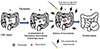 |
Figure 6 Schematic diagram of Thermodox mechanism in CRC: (a) CRC tumour, (b) EPR effect enhances nanoparticle accumulation, (c) Ultrasound-induced local hyperthermia triggers drug release, (d) Tumour undergoes treatment [45]. |
The FDA has approved the liposomal formulation Doxil®, carrying doxorubicin, since the mid-1990s [50]. Doxil is approximately 100 nm in diameter and has much less cardiac and gastrointestinal toxicity. Doxorubicin induces apoptosis by targeting both nuclear and mitochondrial pathways, as shown in Figure 7 [17]. It can cause side effects like skin redness, tenderness, and peeling, which may be painful for some patients [51].
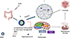 |
Figure 7 Mechanism of action of Doxil on nucleus and mitochondria induces cell death, (a) colorectal cancer, (b) accumulation of Doxorubicin in colorectal tumour, (c) molecular mechanism of Doxorubicin in the nucleus, increase of topoisomerase II, causes more DNA breaking which consequently gives rise to apoptosis but in mitochondria molecular mechanism of Doxorubicin, Fe2+−conjugated Doxorubicin causes Reactive oxygen species production that induces apoptosis and Doxil inhibits the mitochondrial kinases, consequential in apoptosis induction and finally leads to cell death [17]. |
Polymeric nanoparticles
Polymeric nanoparticles have been utilized in the treatment of colon cancer in an encouraging way. They enhance drug delivery, increase bioavailability, and lessen systemic toxicity [52]. These nanoparticles can deliver drugs with precision, making them highly effective for chemotherapy and combination therapies. Several studies have investigated their potential to improve therapeutic outcomes in colon cancer patients. Polymeric nanoparticles improve the delivery of the drug by triggering a response to acidic tumour microenvironments to ensure a targeted release. This pH-dependent action enhances therapy with reduced side effects. Furthermore, functionalized nanoparticles have enabled the targeting of drug delivery via ligand interactions. The carriers are biocompatible carriers composed of poly(lactic-co-glycolide) (PLGA), surface-functionalized with polyethylene glycol (PEG) and ligands such as folate and virus-derived peptides for targeting tumours. The strategy has been experimentally confirmed by the co-loading of camptothecin and curcumin, showing an improved antitumour effect [53]. In contrast, carbohydrate-based polymeric nanoparticles have been applied due to their biodegradability and biocompatibility, offering a favourable approach for colon cancer therapy. Subudhi et al. (2015) [] showed that pectin was discovered to be an efficient carrier for medicine delivery systems aimed at the colon [54]. Eudragit S100-coated Polymeric Nanoparticles (E-CPNs) were formulated to deliver 5-Fluorouracil (5-FU) for colorectal cancer therapy, and their effectiveness was evaluated in both in vivo and in vitro studies [55]. The nanoparticles effectively delayed the drug's release, avoiding premature release in the gastrointestinal tract. This function ensures targeted drug delivery to the colon site [56].
Guar gum (GG) is a promising carrier for colon-specific medication delivery due to its regulated release and microbial degradation [40]. Carboxymethyl guar gum-stabilized nanoparticles were synthesised and tested for dose-dependent cytotoxicity on NHDF cells [57]. Ganesh et al. (2018), showed that 5-FU-loaded guar gum-stabilized nanoparticles exhibited a cytotoxic IC50 value of 1.25 μg/mL against HT-29 cells [58]. Garg et al. (2015), showed that 5-FU-loaded guar gum-chitosan nanoparticles cross-linked with PEG exhibited lower cytotoxicity toward C26 cell lines while sustaining drug release for up to 32 hours. They also demonstrated improved hemocompatibility [59].
Carbon nanotubes (CNT)
Carbon nanotubes (CNTs) are nanomaterials of high promise for drug delivery, gene therapy, and diagnostics. These materials exhibit exceptional optical characteristics, thermal conductivity, and strong chemical stability, which make them highly valuable for biomedical uses. Structurally, they consist of tiny tubular arrangements of carbon atoms forming a honeycomb-like nanostructure. Depending on the number of layers of carbon, CNTs are either single-walled (SWCNTs) or multi-walled (MWCNTs) [44]. Different studies detail the diagnostic and therapeutic use of CNTs in cancer. CNTs with CpG conjugation increase CpG deposition in mouse colon cancer cells and induce NF-κB signalling. This conjugate also significantly suppresses the growth of local xenograft tumours as well as the spread of cancer to the liver metastasis [60]. Fluorescein-modified SWCNT/II-NCC hybrids demonstrated enhanced effectiveness against colon cancer (Caco-2) cells [61]. The functionalised CNTs are more active than their non-functionalized counterparts. SWCNTs conjugated to the C225 antibody are used for targeted therapy against EGFR-expressing CRC cells [41]. The medication is more selective since it targets receptor-mediated endocytosis.
Silica nanoparticles
Silica nanoparticles (SiNPs) possess beehive-like porous morphology with cavity diameters ranging from 50 to 300 nm. Pore diameters range from 2 to 6 nm [62], with a high porous skeleton and good biocompatibility. Low toxicity, pH sensitivity, and easy functionalization render them ideal for drug delivery [63]. In a report on a novel drug delivery system (DDS), colchicine was loaded into spherical mesoporous silica nanoparticles, functionalized with phosphonate groups and coated with a chitosan-glycine–folic acid complex for targeted cancer delivery. The system colchicine-bounded mesoporous silica nanoparticles with phosphonate groups, and coating with a chitosan-glycine–folic acid (MSNsPCOL/CG-FA) achieved complete inhibition of HCT116 colon cancer cells via enhanced intrinsic apoptosis, stronger tubulin inhibition, increased G2/M arrest, and greater suppression of anti-apoptotic proteins compared to free colchicine and 5-FU [64]. All these features render SiNPs highly effective for cancer treatment. A protamine-hyaluronic acid conjugated mesoporous silica NPs enhances activity and loading at tumour locations as synergistic targeted chemo-photothermal therapy [65]. Mesoporous silica nanoparticles (MSNs) offer controlled drug delivery to colorectal cancer (CRC) cells, with enhanced efficacy through gatekeeper modifications such as stimuli-responsive systems [66]. Fan et al. (2022) developed a dual-targeting, pH- and redox-sensitive formulation by conjugating a CEA/CD44-targeting nanobody (11C12) and hyaluronic acid to doxorubicin-loaded MSNs (DOX@MSNs-HA-11C12). In vitro cytotoxicity studies on LoVo CRC cells demonstrated the formulation's good biocompatibility and safety, along with significantly increased apoptosis. The study concluded that this dual-targeted nanocarrier enhances the precision and therapeutic efficiency of drug delivery to CRC cells [67].
Nanoemulsion system
A nanoemulsion, a system composed of oil, water, and a surfactant, is a translucent colloidal solution. These systems exhibit advantages such as low toxicity, high stability, thermosensitivity, pH responsiveness and improved efficacy [68, 69]. Anti-angiogenic agents, commonly used for CRC, often cause toxicity, lack target selectivity, and struggle with tumour penetration. Nanoemulsions help overcome these challenges by improving drug uptake and enabling deeper penetration into highly vascularized tumour tissues [70]. Their hydrophobic core facilitates efficient encapsulation of water-insoluble drugs to improve therapeutic effects. Antibody conjugation facilitates targeted delivery, while PEGylation improves circulation, retention and creates a protective hydrophilic layer on the nanoparticle surface, reducing protein corona formation and immune evasion [71, 72]. Huang et al. (2015) examined the synergic effect of a nanoemulsion formulated with Tween 80 as the emulsifier, incorporating lycopene (LP) and gold nanoparticles (AN). It was tested on the HT-29 colon cancer cell line, exhibiting an 80 % reduction in cell survival at low concentrations of AN (0.16 ppm) and LP (0.4 μM) that is significantly higher than the 50–60% caused by the co-treatment of AN and LP alone. Mechanistic studies indicated that the nanoemulsion altered the expression of apoptosis and signalling proteins by suppressing procaspase-8, procaspase-3, procaspase-9, PARP-1 and Bcl-2 and increasing Bax expression. Additionally, nanoemulsion inhibited cell migration and invasion through the upregulation of E-cadherin and downregulation of Akt and NFκB signaling pathways, highlighting its potential as a promising therapeutic strategy for colorectal cancer [72].
Case study
Formulation and Characterization of Doxycycline-Loaded Polymeric Nanoparticles for Testing Antitumor/Antiangiogenic Action in Experimental Colon Cancer in Mice.
In a study on artificially induced colon cancer using 1,2-dimethylhydrazine, Doxycycline-Loaded Polymeric Nanoparticles (DOX-PRNP) were injected subcutaneously to overcome limitations of free doxycycline (DOX), such as systemic toxicity, rapid clearance, insufficient tumour-site concentration, and mitochondrial biogenesis inhibition that affects healthy cells. Treatment with DOX-PRNP resulted in better histopathological findings in the colon, including fewer malignant alterations and decreased tumour burden. This was evidenced by significantly lower scores for crypt distortion, hyperplasia, loss of goblet cells, and dysplasia. Additionally, the nanoparticles significantly reduced angiogenic markers VEGF and CD31 in tumour tissues, indicating the nanocarrier system enhances the therapeutic efficacy of doxycycline and presents a promising approach for more effective colon cancer treatment [73].
Conclusion
The development of nanoparticles for targeted drug delivery has been a major breakthrough in treating patients with CRC, as they allow for localized, controlled release of anticancer agents with reduced systemic side effects. This study illustrates the potential of diverse nanocarriers, such as liposomes, polymeric nanoparticles, carbon nanotubes, silica nanoparticles, and nanoemulsions, for surpassing biological barriers, increasing drug accumulation in tumour tissues, and consequently improving therapy. Clinically, US FDA-approved formulations such as Doxil and investigational agents like CPX-1 and LE-SN38 demonstrate the growing potential of nanomedicine in CRC treatment. Although challenges remain in optimizing delivery strategies and identifying highly specific molecular biomarkers, continuous research in nanobioconjugation and multifunctional delivery systems is paving the way for more effective, personalized, and safer therapeutic approaches.
Future prospective
The advancement of nanoparticle-mediated targeted therapies presents a promising future for colorectal cancer (CRC) treatment, particularly through nanobioconjugation approaches that enable precise drug delivery with reduced systemic toxicity. Functionalized nanoparticles offer controlled release, enhanced therapeutic efficacy, and reduced side effects, making them ideal candidates for advanced treatment modalities. Emphasis should now shift toward refining these systems for better pharmacokinetics and compatibility with combination therapies. To ensure clinical translation, it is essential to conduct extensive clinical trials that assess safety and efficacy across diverse patient populations. Ultimately, gaining regulatory approvals such as from the FDA will be crucial in integrating these nanomedicine strategies into standard CRC care, driving a shift toward safer and more effective therapeutic options.
Funding
This review did not receive any specific funding.
Conflicts of interest
The authors declare no conflict of interest.
Data availability statement
Data will be made available on request.
Author contribution statement
Credit authorship contribution statement Siddananjaiah Karthik: Conceptualization, Methodology, Investigation, Writing – original draft. Soumya V Menon: Review & editing, Validation. Shankar Sumanth- Investigation, Figures. Shivamogga Natesh Ananya: Methodplogy, Figures.
Ethics approval
Ethical approval was not required.
Acknowledgments
The authors highly acknowledge the guidance and support facility provided by Dr. Soumya V Menon, Department of Chemistry and Biochemistry, School of Sciences, JAIN (Deemed-to-be University), JC Road, Sudhama Nagar, Bengaluru, Karnataka for the preparation of the manuscript.
References
- Nussbaum YI, Manjunath Y, Suvilesh KN, et al. Current and prospective methods for assessing anti-tumor immunity in colorectal cancer. Int J Mol Sci. 2021;22:4802. https://doi.org/10.3390/ijms22094802. [Google Scholar]
- Shivshankar S, Patil PS, Deodhar K, et al. Epidemiology of colorectal cancer: A review with special emphasis on India. Indian J Gastroenterol. 2025;44:142–153. https://doi.org/10.1007/s12664-024-01726-8. [Google Scholar]
- CRI New York, 2025 – https://www.cancerresearch.org/blog/colorectal-cancer-awareness-month#_ftn1. [Google Scholar]
- Hamzehzadeh L, Imanparast A, Tajbakhsh A, et al. New approaches to use nanoparticles for treatment of colorectal cancer; a brief review. Nanomed Res J. Tehran University of Medical Sciences 2016a;1:59–68. https://doi.org/10.7508/NMRJ.2016.02.001. [Google Scholar]
- WHO, 2023 – https://www.who.int/news-room/fact-sheets/detail/colorectal-cancer. [Google Scholar]
- Banerjee A, Pathak S, Subramanium VD, et al. Strategies for targeted drug delivery in treatment of colon cancer: current trends and future perspectives. Drug Discovery Today. 2017;22:1224–1232. https://doi.org/10.1016/j.drudis.2017.05.006. [Google Scholar]
- Patel K, Jain P, Rajput PK, et al. Human serum albumin-based propulsive Piperlongumine-loaded nanoparticles: Formulation development, characterization and anti-cancer study. Colloids Surf A: Physicochem Eng Aspects. 2022;652:129738. https://doi.org/10.1016/j.colsurfa.2022.129738. [Google Scholar]
- Reddy RG, Suvarna DVAH, Kumar RVT. Colorectal cancer in India: An audit from a tertiary care centre. Res Article J MAR Gastroenterol. 2023;2:1. www.medicalandresearch.com. https://www.medicalandresearch.com/previous_view/1146. [Google Scholar]
- Ghazarian H, Idoni B, Oppenheimer SB. A glycobiology review: Carbohydrates, lectins and implications in cancer therapeutics. Acta Histochemica. 2011;113:236–247. https://doi.org/10.1016/j.acthis.2010.02.004. [Google Scholar]
- Castro E, Kumar A. Nanoparticles in drug delivery systems. In Nanomedicine in drug delivery . CRC Press;2013. p. 1–22. https://doi.org/10.1201/b14802. [Google Scholar]
- Davis ME, Chen Z, Shin DM. Nanoparticle therapeutics: An emerging treatment modality for cancer. In Nature reviews drug discovery (Vol. 7). Nature Publishing Group; 2008. p. 771–782. https://doi.org/10.1038/nrd2614. [Google Scholar]
- Cepero A, Luque C, Cabeza L, et al. Antibody-functionalized nanoformulations for targeted therapy of colorectal cancer: A systematic review. Int J Nanomed. 2022;17:5065–5080. Dove Medical Press Ltd. https://doi.org/10.2147/IJN.S368814. [Google Scholar]
- Wang AZ, Langer R, Farokhzad OC. Nanoparticle delivery of cancer drugs. Annu Rev Med. 2012;63:185–198. https://doi.org/10.1146/annurev-med-040210-162544. [Google Scholar]
- Menon DS, Abira R, Gokarn S, et al. Multifaceted peptide-based nanoparticles for diagnosis of brain tumors: Recent advances and future outlooks. J Biotech Biomed. 2025;8:59–71. https://doi.org/10.26502/jbb.2642-91280179. [Google Scholar]
- Amaral M, Hamad H, Menon DS, et al. The effect of cynaropicrin, a sesquiterpene lactone, on the migratory properties of triple-negative breast cancer cells and the underlying mechanisms. Avicenna J Phytomed. 2025;16:66–77. https://doi.org/10.22038/ajp.2025.25894. [Google Scholar]
- Al-Joufi FA, Setia A, Salem-Bekhit MM, et al. Molecular pathogenesis of colorectal cancer with an emphasis on recent advances in biomarkers, as well as nanotechnology-based diagnostic and therapeutic approaches. Nanomaterials. 2022;12(1):169. https://doi.org/10.3390/nano12010169. [Google Scholar]
- Narayana S, Gowda BHJ, Hani U, et al. Inorganic nanoparticle-based treatment approaches for colorectal cancer: recent advancements and challenges. J Nanobiotechnol. 2024;22:427. https://doi.org/10.1186/s12951-024-02701-3. [Google Scholar]
- Robles AI, Traverso G, Zhang M, et al. Whole-exome sequencing analyses of inflammatory bowel disease-associated colorectal cancers. Gastroenterology. 2016;150:931–943. https://doi.org/10.1053/j.gastro.2015.12.036. [Google Scholar]
- Swiderska M, Choromańska B, Dąbrowska E, et al. The diagnostics of colorectal cancer. Contemp Oncol (Poznan, Poland). 2014;18:1–6. https://doi.org/10.5114/wo.2013.39995. [Google Scholar]
- Levin B, Lieberman DA, McFarland B, et al. Screening and surveillance for the early detection of colorectal cancer and adenomatous polyps, 2008: A joint guideline from the american cancer society, the US multi-society task force on colorectal cancer, and the American college of radiology. CA: A Cancer J Clin. 2008;58:130–160. https://doi.org/10.3322/ca.2007.0018. [Google Scholar]
- Szajda SD, Borzym-Kluczyk M, Snarska J, et al. N-acetyl-β-D-hexosaminidase and its isoenzymes A and B in blood serum and urine, as a potential colon cancer markers. Hepato-Gastroenterol. 2009;56:1287–1298. [Google Scholar]
- Zhang YL, Zhang ZS, Wu BP, et al. Early diagnosis for colorectal cancer in China. World J Gastroenterol. 2002;8(1):21–25. https://doi.org/10.3748/wjg.v8.i1.21. [Google Scholar]
- Tang X, Liang Y, Feng X, et al. Co-delivery of docetaxel and Poloxamer 235 by PLGA-TPGS nanoparticles for breast cancer treatment. Mater Sci Eng C. 2015;49:348–355. https://doi.org/10.1016/j.msec.2015.01.033. [Google Scholar]
- Ades S. Adjuvant chemotherapy for colon cancer in the elderly: Moving from evidence to practice. Oncology. 2009;23:162–167. [Google Scholar]
- Kumar A, Gautam V, Sandhu A, et al. Current and emerging therapeutic approaches for colorectal cancer: A comprehensive review. World J Gastrointest Surg. 2023; 15(4):495–519. https://doi.org/10.4240/wjgs.v15.i4.495. [Google Scholar]
- Zhang YR, Zhu HR, Li HR, et al. Trends in nanomedicine for colorectal cancer treatment: Bibliometric and visualization analysis (2010–2024). World Journal of Gastrointest Oncol. 2025;17(4):102438. https://doi.org/10.4251/wjgo.v17.i4.102438. [Google Scholar]
- Overman MJ, Lonardi S, Wong KYM, et al. Durable clinical benefit with nivolumab plus ipilimumab in DNA mismatch repair-deficient/microsatellite instability-high metastatic colorectal cancer. J Clin Oncol. 2018;36:773–779. https://doi.org/10.1200/JCO.2017.76.9901. [Google Scholar]
- Viralippurath Ashraf J, Sasidharan Nair V, Saleh R, et al. Role of circular RNAs in colorectal tumor microenvironment. In Biomedicine and pharmacotherapy (Vol. 137). Elsevier Masson s.r.l; 2021. https://doi.org/10.1016/j.biopha.2021.111351. [Google Scholar]
- Raju GSR, Pavitra E, Varaprasad GL, et al. Nanoparticles mediated tumor microenvironment modulation: current advances and applications. J Nanobiotechnol. 2022;20:274. https://doi.org/10.1186/s12951-022-01476-9. [Google Scholar]
- Yang M, Li J, Gu P, et al. The application of nanoparticles in cancer immunotherapy: Targeting tumor microenvironment. Bioact Mater. 2021;6(7):1973–1987. https://doi.org/1s0.1016/j.bioactmat.2020.12.010. [Google Scholar]
- Pandit S, Dutta D, Nie S. Active transcytosis and new opportunities for cancer nanomedicine. Nat Mater. 2020;19(5):478–480. https://doi.org/10.1038/s41563-020-0672-1. [Google Scholar]
- Maeda H. Toward a full understanding of the EPR effect in primary and metastatic tumors as well as issues related to its heterogeneity. Adv Drug Delivery Rev. 2015;91:3–6. https://doi.org/10.1016/j.addr.2015.01.002. [Google Scholar]
- Li X, Hu Y, Zhang X, et al. Transvascular transport of nanocarriers for tumor delivery. Nat Commun. 2024;15:8172. https://doi.org/10.1038/s41467-024-52416-0. [Google Scholar]
- Hasan M, Evett C, Burton J. Advances in nanoparticle-based targeted drug delivery systems for colorectal cancer therapy: A review. arXiv. 2024. https://doi.org/10.48550/arXiv.2409.05222. [Google Scholar]
- Rodrigues P, Thiab A, Jabir M, et al. Combination therapies and novel delivery systems: A new frontier in overcoming TRAIL resistance in gastric cancer. Naunyn-Schmiedeberg's Archives of Pharmacology. 2025;398:13581–13603. https://doi.org/10.1007/s00210-025-04208-6. [Google Scholar]
- Carvalho MR, Carvalho CR, Maia FR, et al. Peptide-modified dendrimer nanoparticles for targeted therapy of colorectal cancer. Adv Ther. 2019;2:1900132. https://doi.org/10.1002/adtp.201900132. [Google Scholar]
- Briolay T, Petithomme T, Fouet M, et al. Delivery of cancer therapies by synthetic and bio-inspired nanovectors. In Molecular cancer (Vol. 20). BioMed Central Ltd; 2021. https://doi.org/10.1186/s12943-021-01346-2. [Google Scholar]
- Choi KY, Jeon EJ, Yoon HY, et al. Theranostic nanoparticles based on PEGylated hyaluronic acid for the diagnosis, therapy and monitoring of colon cancer. Biomaterials. 2012;33(26):6186–6193. https://doi.org/10.1016/j.biomaterials.2012.05.029. [Google Scholar]
- Alkhader E, Roberts CJ, Rosli R, et al. Pharmacokinetic and anti-colon cancer properties of curcumin-containing chitosan-pectinate composite nanoparticles. J Biomater Sci Polymer Ed. 2018;29(18):2281–2298. https://doi.org/10.1080/09205063.2018.1541500. [Google Scholar]
- Baylisss CE, Houston AP. Characterization of plant polysaccharide- and mucin-fermenting anaerobic bacteria from human feces. Appl Environ Microbiol. 1984;48:626–632. https://doi.org/10.1128/aem.48.3.626-632.1984. [Google Scholar]
- Lee PC, Chiou YC, Wong JM, et al. Targeting colorectal cancer cells with single-walled carbon nanotubes conjugated to anticancer agent SN-38 and EGFR antibody. Biomaterials. 2013;34:8756–8765. https://doi.org/10.1016/j.biomaterials.2013.07.067. [Google Scholar]
- Venturini J, Chakraborty A, Baysal MA et al. Developments in nanotechnology approaches for the treatment of solid tumors. Exp Hematol Oncol. 2025;14:76. https://doi.org/10.1186/s40164-025-00656-1. [Google Scholar]
- Batist G, Sawyer M, Gabrail N, et al. A multicenter, phase II study of CPX-1 liposome injection in patients (pts) with advanced colorectal cancer (CRC). J Clin Oncol. 2008;26:4108–4108. https://doi.org/10.1200/jco.2008.26.15_suppl.4108. [Google Scholar]
- Gogoi P, Kaur G, Singh NK. Nanotechnology for colorectal cancer detection and treatment. World J Gastroenterol. 2022;28:6497–6511. Baishideng Publishing Group Inc. https://doi.org/10.3748/wjg.v28.i46.6497. [Google Scholar]
- Dou Y, Hynynen K, Allen C. To heat or not to heat: Challenges with clinical translation of thermosensitive liposomes. J Controlled Release. 2017;249: 63–73. https://doi.org/10.1016/j.jconrel.2017.01.025. [Google Scholar]
- Yin L, Li H, Shi L, et al. Research advances in nanomedicine applied to the systemic treatment of colorectal cancer. Int J Cancer. 2023;152:807–821. [Google Scholar]
- Ter Haar G; Shaw A. Requirements for measurement standards in high intensity ultrasound (HIFU) fields. NPL Rep. 2006;15:DQL-AC 015. [Google Scholar]
- Arulpragasam AR, van ’t Wout-Frank M, Barredo J, et al. Low intensity focused ultrasound for non-invasive and reversible deep brain neuromodulation – a paradigm shift in psychiatric research. Front Psychiatry. 2022;13:825802. [Google Scholar]
- Farooqi MA, Mahnoor M, Delgado KM, et al. Focused ultrasound as targeted therapy for colorectal cancer: A comprehensive review. Gastrointest Disord. 2024;6(2):380–401. https://doi.org/10.3390/gidisord6020026. [Google Scholar]
- Barenholz Y. Doxil® - The first FDA-approved nano-drug: Lessons learned J Controlled Release. 2012;160:117–134. https://doi.org/10.1016/j.jconrel.2012.03.020. [Google Scholar]
- Campos S. Liposomal anthracyclines: Adjuvant and neoadjuvant therapy for breast cancer. Oncologist. 2003;8:10–16. https://doi.org/10.1634/theoncologist.8-suppl_2-10. [Google Scholar]
- Udaipuria N, Bhattacharya S. Novel carbohydrate polymer-based systems for precise drug delivery in colon cancer: improving treatment effectiveness with intelligent biodegradable materials. Biopolymers. 2024;116(1):e23632. https://doi.org/10.1002/bip.23632. [Google Scholar]
- Sun Y, Li B, Cao Q, et al. Targeting cancer stem cells with polymer nanoparticles for gastrointestinal cancer treatment. Stem Cell Res Ther. 2022;13:489. https://doi.org/10.1186/s13287-022-03180-9. [Google Scholar]
- Subudhi MB, Jain A, Jain A, et al. Eudragit S100 coated citrus pectin nanoparticles for colon targeting of 5-fluorouracil. Mater (Basel, Switzerland). 2015;8(3):832–849. https://doi.org/10.3390/ma8030832. [Google Scholar]
- Tummala S, Satish Kumar MN, Prakash A. Formulation and characterization of 5-Fluorouracil enteric coated nanoparticles for sustained and localized release in treating colorectal cancer. Saudi Pharm J. 2015;23:308–314. https://doi.org/10.1016/j.jsps.2014.11.010. [Google Scholar]
- Asghar LFA, Chandran S. Design and evaluation of matrices of Eudragit with polycarbophil and carbopol for colon-specific delivery. J Drug Targeting. 2008;16:741–757. https://doi.org/10.1080/10611860802473345. [Google Scholar]
- Dodi G, Pala A, Barbu E, et al. Carboxymethyl guar gum nanoparticles for drug delivery applications: Preparation and preliminary in-vitro investigations. Mater Sci Eng C. 2016;63:628–636. https://doi.org/10.1016/j.msec.2016.03.032. [Google Scholar]
- Ganeshkumar M, Janani M, Ponrasu T, et al. Guar gum stabilized gold nanoparticles for colon cancer treatment. Clin Oncol. 2018;3:1539. [Google Scholar]
- Garg S, Shukla A, Garg A. Design and characterization of Guar gum coated chitosan nanoparticles for delivery of 5-fluorouracil for effective treatment of colon cancer. J Med Pharm Allied Sci. 2015;45:28–45. [Google Scholar]
- Jin H, Gao S, Song D, et al. Intratumorally CpG immunotherapy with carbon nanotubes inhibits local tumor growth and liver metastasis by suppressing the epithelial-mesenchymal transition of colon cancer cells. Anti-Cancer Drugs. 2021;32:278–285. https://doi.org/10.1097/CAD.0000000000001000. [Google Scholar]
- González-Domínguez JM, Grasa L, Frontiñán-Rubio J, et al. Intrinsic and selective activity of functionalized carbon nanotube/nanocellulose platforms against colon cancer cells. Colloids Surf B: Biointerfaces. 2022;212:112363. https://doi.org/10.1016/j.colsurfb.2022.112363. [Google Scholar]
- Stang J, Haynes M, Carson P, et al. A preclinical system prototype for focused microwave thermal therapy of the breast. IEEE Trans Biomed Eng. 2012a;59:2431–2438. https://doi.org/10.1109/TBME.2012.2199492. [Google Scholar]
- Bharti C, Nagaich U, Pal AK, et al. Nanopartículas de sílice mesoporosa en sistemas de administración de fármacos: Una revisión. Revista Internacional de Investigación Farmacéutica. 2015;5:124. [Google Scholar]
- AbouAitah K, Hassan HA, Swiderska-Sroda A, et al. Targeted nano-drug delivery of colchicine against colon cancer cells by means of mesoporous silica nanoparticles. Cancers. 2020;12(1):144. https://doi.org/10.3390/cancers12010144. [Google Scholar]
- Chen C, Tang W, Jiang D, et al. Hyaluronic acid conjugated polydopamine functionalized mesoporous silica nanoparticles for synergistic targeted chemo-photothermal therapy. Nanoscale. 2019;11:11012–11024. https://doi.org/10.1039/c9nr01385g. [Google Scholar]
- Cheng W, Nie J, Xu L, et al. PH-sensitive delivery vehicle based on folic acid-conjugated polydopamine-modified mesoporous silica nanoparticles for targeted cancer therapy. ACS Appl Mater Interfaces. 2017;9:18462–18473. https://doi.org/10.1021/acsami.7b02457. [Google Scholar]
- Fan X, Wang T, Han M, et al. Dual CEA/CD44 targeting to colorectal cancer cells using nanobody-conjugated hyaluronic acid-modified mesoporous silica nanoparticles with pH- and redox-sensitivity. Mater Adv. 2022;3(14):4707–4717. https://doi.org/10.1039/d2ma00082b. [Google Scholar]
- Silva R, Ferreira H, Cavaco-Paulo A. Sonoproduction of liposomes and protein particles as templates for delivery purposes. Biomacromolecules. 2011;12:3353–3368. https://doi.org/10.1021/bm200658b. [Google Scholar]
- Dezfuli AAZ, Abu-Elghait M, Salem SS. Recent insights into nanotechnology in colorectal cancer. In Applied biochemistry and biotechnology (Vol. 196). Springer; 2024. p. 4457–4471. https://doi.org/10.1007/s12010-023-04696-3. [Google Scholar]
- Ganta S, Talekar M, Singh A, et al. Nanoemulsions in translational research – Opportunities and challenges in targeted cancer therapy. In AAPS PharmSciTech (Vol. 15). New York LLC: Springer; 2014. p. 694–708. https://doi.org/10.1208/s12249-014-0088-9. [Google Scholar]
- Sánchez-López E, Guerra M, Dias-Ferreira J, et al. Current applications of nanoemulsions in cancer therapeutics. Nanomater (Basel, Switzerland). 2019;9:821. https://doi.org/10.3390/nano9060821. [Google Scholar]
- Huang RF, Wei YJ, Inbaraj BS, et al. Inhibition of colon cancer cell growth by nanoemulsion carrying gold nanoparticles and lycopene. Int J Nanomed. 2015;10:2823–2846. https://doi.org/10.2147/IJN.S79107. [Google Scholar]
- Alshaman R, Alattar A, El-Sayed RM, et al. Formulation and characterization of doxycycline-loaded polymeric nanoparticles for testing antitumor/antiangiogenic action in experimental colon cancer in mice. Nanomaterials. 2022;12:857. https://doi.org/10.3390/nano12050857. [Google Scholar]
Cite this article as: Siddananjaiah K, Shankar S, Natesh AS, & Menon SV. Multifunctional nanoparticles in colorectal cancer therapy: Advances in targeted drug delivery systems. Visualized Cancer Medicine. 2026; 7, 3. https://doi.org/10.1051/vcm/2026002.
All Tables
Different types of nanocarriers in drug delivery system for colorectal carcinoma treatment.
All Figures
|
Figure 1 Development of colorectal cancer: Initiation, promotion, progression, local progression, metastasis and common metastatic sites [14]. |
| In the text | |
|
Figure 2 Schematic representation of the tumour microenvironment and its cellular composition of colorectal cancer [17, 28]. |
| In the text | |
|
Figure 3 Pleiotropic mechanisms in the tumour microenvironment (TME). (a) Drug loaded NPs can inhibit the hypoxic tumor vasculature of colorectal cancer by targeting and reprogramming the multiple mechanisms: i) Myeloid-Derived Suppressor Cells (MDSCs) and T-regulatory cells (Tregs) inhibit cytotoxic T-lymphocytes (CTL) and drive tumour growth. ii) The conversion of M1 macrophages to M2 macrophages also leads to tumor growth. iii) TGFβ-induced fibroblasts activate Cancer-Associated Fibroblasts (CAFs), which promote tumor progression. iv) VEGF also stimulates abnormal tumour vasculature and cancer development. Drug loaded NPs can target all of these pathways to suppress tumour growth. NPs can reprogram M2 macrophages into M1 phenotypes, which reactivate the immune response and inhibit tumour growth. Drug loaded NPs can also activate mature DCs, leading to the activation of B cells through T cells and the production of antibodies that can target and eradicate tumour cells. These NP-stimulated pathways can thus overcome the inhibitory effects exerted by MDSCs on mature dendritic cells (DCs) and the inhibitory effects of Tregs on INF-γ, thus killing tumour cells. Additionally, NPs loaded with oxygen can inhibit the hypoxic tumour vasculature by inducing oxygen, thus reversing hypoxia and limiting tumour growth [29]. (b) Schematic representation of how drug-loaded nanoparticles modulate tumor-associated macrophages (TAMs) under hypoxic conditions. NPs inhibit TAM2 and promote TAM1 polarization, leading to increased IL-12 and decreased IL-10 production, which suppresses tumour vasculature and enhances anti-tumor immunity. Drug-loaded NPs can inhibit the conversion of TAM1 to TAM2 and reduce tumour growth. SiRNA-loaded NPs can also suppress TAM2 activity [30]. |
| In the text | |
|
Figure 4 Normal microvessels with thick endothelial cells and tumour microvessels with leaky endothelial gaps due to overexpression of VEGFR (a) [33]. NPs are trapped in the subendothelial void by basement membrane surrounding vessels, forming a perivascular NPs pool that hinders their extravasation (b). Collagen hydrolase-mediated basement membrane destruction is used to increase NP extravasation (c) [34]. |
| In the text | |
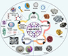 |
Figure 5 Different types of nanoparticles for cancer therapy [37]. |
| In the text | |
|
Figure 6 Schematic diagram of Thermodox mechanism in CRC: (a) CRC tumour, (b) EPR effect enhances nanoparticle accumulation, (c) Ultrasound-induced local hyperthermia triggers drug release, (d) Tumour undergoes treatment [45]. |
| In the text | |
|
Figure 7 Mechanism of action of Doxil on nucleus and mitochondria induces cell death, (a) colorectal cancer, (b) accumulation of Doxorubicin in colorectal tumour, (c) molecular mechanism of Doxorubicin in the nucleus, increase of topoisomerase II, causes more DNA breaking which consequently gives rise to apoptosis but in mitochondria molecular mechanism of Doxorubicin, Fe2+−conjugated Doxorubicin causes Reactive oxygen species production that induces apoptosis and Doxil inhibits the mitochondrial kinases, consequential in apoptosis induction and finally leads to cell death [17]. |
| In the text | |
Current usage metrics show cumulative count of Article Views (full-text article views including HTML views, PDF and ePub downloads, according to the available data) and Abstracts Views on Vision4Press platform.
Data correspond to usage on the plateform after 2015. The current usage metrics is available 48-96 hours after online publication and is updated daily on week days.
Initial download of the metrics may take a while.