| Issue |
Vis Cancer Med
Volume 7, 2026
|
|
|---|---|---|
| Article Number | E1 | |
| Number of page(s) | 3 | |
| DOI | https://doi.org/10.1051/vcm/2025016 | |
| Published online | 13 March 2026 | |
Editorial
Interwoven control of histone lactylation: metabolic enzymes LDHA/ACSS2 couple with epigenetic writer KAT2A
1
Zhejiang Key Laboratory of Pancreatic Disease, The First Affiliated Hospital, Zhejiang Key Laboratory of Frontier Medical Research on Cancer Metabolism, Institute of Translational Medicine, Zhejiang University School of Medicine, Hangzhou, Zhejiang 310029, PR China
2
Institute of Fundamental and Transdisciplinary Research, Cancer Center, Zhejiang University, Hangzhou, Zhejiang 310029, PR China
3
The Center for Translational Research, The Second Affiliated Hospital and Institute of Translational Medicine, Zhejiang University, Hangzhou, Zhejiang 310029, PR China
* Corresponding author: This email address is being protected from spambots. You need JavaScript enabled to view it.
Received:
22
May
2025
Accepted:
10
December
2025
Abstract
Histone lactylation is critically involved in the regulation of gene expression and the modulation of diverse cellular processes in both normal and tumor cells. A recent study by Zhu et al. [Cell Metab. 37, 361–376] demonstrated that ACSS2 functions as a bona fide lactyl-CoA synthetase, converting lactate into lactyl-CoA. In response to elevated aerobic glycolysis and EGFR activation, ERK-mediated phosphorylation drives the nuclear translocation of ACSS2, promoting the formation of the ACSS2–KAT2A complex. KAT2A utilizes ACSS2-derived lactyl-CoA and acts as a lactyltransferase to promote histone lactylation, gene expression, tumor growth, and immune evasion.
Key words: Histone / Lactylation / ACSS2 / KAT2A
These authors have contributed equally to the article.
© The Authors, published by EDP Sciences, 2026
 This is an Open Access article distributed under the terms of the Creative Commons Attribution License (https://creativecommons.org/licenses/by/4.0), which permits unrestricted use, distribution, and reproduction in any medium, provided the original work is properly cited.
The Warburg effect, which describes the tendency of tumor cells to generate energy through aerobic glycolysis even under well-oxygenated conditions, is considered a key metabolic feature of tumors [1–3]. This metabolic reprogramming enhances the production of lactate, which can be converted into lactyl-coenzyme A (lactyl-CoA), the donor molecule for both enzymatic and non-enzymatic protein lactylation [4]. Histone H3 can be lactylated at lysines 14 (K14) and 18 (K18), leading to homeostatic gene transcription [4]. However, lactyl-CoA synthetases responsible for generating lactyl-CoA have not yet been identified.
A recent seminal finding by Zhu et al. demonstrates for the first time that acetyl-CoA synthetase 2 (ACSS2) is a bona fide lactyl-CoA synthetase, forming a complex with lactate dehydrogenase A (LDHA) and the acetyltransferase KAT2A to coordinately promote KAT2A-mediated histone H3 lactylation and gene expression for tumor growth and immune evasion [4]. In response to hypoxia, lactate stimulation, KRAS G12C mutation, and activation of epidermal growth factor receptor (EGFR), platelet-derived growth factor receptor (PDGFR), and fibroblast growth factor receptor (FGFR), glioblastoma (GBM) cells – unlike normal astrocytes – exhibited markedly increased lactylation of histone H3 at lysines 14 (K14) and 18 (K18). However, incubation of purified histone H3 with 1–100 nM lactyl-CoA, a range corresponding to nuclear concentrations observed in EGF-treated primary GBM cells, did not induce H3 lactylation, suggesting the requirement of a histone lactyltransferase [4].
To identify the responsible lactyltransferase, known histone acyltransferases – including P300, CREB-binding protein (CBP), and KAT2A [5, 6] – were individually depleted. Only KAT2A depletion led to a significant reduction in EGF-induced H3 lactylation. Consistently, in vitro assays showed that purified wild-type (WT) KAT2A catalyzed lactylation of histone H3 at K14 and K18, while an enzymatically inactive KAT2A mutant lacked this activity [4]. A co-crystal structure of KAT2A bound to lactyl-CoA (Video 1) reveals that the side chain of R533 extends laterally to form a hydrogen bond with the hydroxyl group of the lactyl moiety. The R533A mutation impaired both the binding and catalytic activity of KAT2A toward lactyl-CoA, without affecting its activity toward other acyl-CoAs. Furthermore, this lactyltransferase-deficient KAT2A mutant significantly reduced EGF-induced H3 K14/K18 lactylation in GBM cells [4]. Collectively, these findings identify KAT2A as a bona fide histone lactyltransferase.
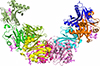 |
Video 1 The structure of KAT2A in complex with lactyl-CoA. The indicated structure (PDB ID: 8E6O) represents the domain crystal structure of the human histone acetyltransferase KAT2A. It contains eight chains (shown in different colors), derived from Homo sapiens, and was solved by X-ray diffraction at 2.37 Å resolution. The structure includes a bound ligand, lactyl-CoA (highlighted in purple in the video), which occupies a deep cleft within KAT2A. The lactyl moiety of lactyl-CoA extends toward the terminus of flexible loop 3, illustrating its specific positioning within the catalytic pocket. https://vcm.edpsciences.org/10.1051/vcm/2025016/olm |
To identify the enzyme responsible for lactyl-CoA synthesis, KAT2A was immunoprecipitated from EGF-treated GBM cells and analyzed by mass spectrometry. Moreover, molecular docking revealed that ACSS2 interacts with KAT2A via residues E299 and E301 of ACSS2. Inhibition of EGFR downstream signaling pathways identified ERK1/2 as a key regulator of this interaction. Specifically, ERK1/2 was found to interact with ACSS2 at residues L432 and V434 and to phosphorylate ACSS2 at serine 267 (S267) [4]. The phosphorylated S267–P268 motif undergoes cis–trans isomerization by the peptidyl-prolyl isomerase PIN1 (Protein Interacting with Never in Mitosis A-1) [4, 7, 8]. This conformational change exposes the nuclear localization signal (NLS) of ACSS2 (R664/R665), facilitating its recognition by importin α5 and promoting nuclear translocation [4].
Although ACSS2 is conventionally known to convert acetate into acetyl-CoA [4], in vitro assays demonstrated its ability to bind lactate. Notably, purified ACSS2 was shown to catalyze the conversion of lactate and CoA into lactyl-CoA, as confirmed by mass spectrometry [4]. Furthermore, incubation of purified KAT2A and ACSS2 in lactate-containing conditions led to ACSS2-dependent histone H3 K14/K18 lactylation, supporting a functional role for ACSS2-coupled KAT2A in histone lactylation. Importantly, EGF treatment induced the formation of a nuclear complex comprising LDHA, ACSS2, and KAT2A in GBM cells [4]. These findings suggest that this complex facilitates the use of LDHA-generated nuclear lactate by ACSS2 for lactyl-CoA production, enabling KAT2A-mediated histone lactylation. These results reveal that ACSS2 is a bona fide lactyl-CoA synthetase.
To evaluate the impact of ACSS2/KAT2A-mediated histone lactylation on gene expression, chromatin immunoprecipitation sequencing (ChIP-seq) was performed in GBM cells. H3K18 lactylation and KAT2A binding co-occurred at 2,028 annotated gene promoter regions. Additionally, approximately 52.5% of H3K18 lactylation sites overlapped with ACSS2-binding regions. These modifications and protein associations were enriched in genes involved in the Wnt, NF-κB, PD-L1, and glycolysis pathways, promoting the expression of WNT2, RELA, and CD274 [4].
Blocking the nuclear translocation of ACSS2 or the binding of KAT2A to lactyl-CoA or ACSS2 inhibited brain tumor growth and prolonged mouse survival. These interventions also enhanced CD8+ T-cell infiltration and granzyme B expression in tumor tissues while reducing the expression of Wnt-2, p65, and PD-L1. Notably, treatment with a peptide that disrupts the ACSS2–KAT2A interaction significantly reduced tumor volume, extended survival, and synergized with anti-PD-1 antibody therapy to enhance anti-tumor effects [4]. Analysis of human GBM specimens revealed a positive correlation between ACSS2 S267 phosphorylation and H3K18 lactylation levels, both of which were associated with elevated PD-L1 expression, reduced CD8+ T-cell infiltration, and poor patient prognosis [4]. Collectively, these findings demonstrate that the ACSS2/KAT2A-axis-dependent histone H3 lactylation facilitates tumor immune evasion and promotes brain tumor progression. Thus, disrupting this pathway suppresses tumor growth and enhances the efficacy of immune checkpoint blockade therapy.
This study demonstrates for the first time that ACSS2 functions as a bona fide lactyl-CoA synthetase. A recent study reports that nuclear GTP-specific succinyl-CoA synthetase (GTPSCS) acts as a lactyl-CoA synthetase that promotes histone lactylation and gliomagenesis [9], suggesting that histone lactylation can be regulated by distinct lactyl-CoA synthetases across different tumor types and in response to diverse oncogenic signaling pathways. KAT2A is frequently overexpressed rather than mutated in tumors [10, 11]. KAT2A acts as a histone lactyltransferase when in complex with ACSS2 and LDHA, thereby regulating tumor immune evasion and promoting tumor growth. These findings suggest that KAT2A promotes tumorigenesis through diverse mechanisms dictated by its binding partners. The discovery that the Warburg effect can directly influence chromatin structure and modulate immune escape mechanisms highlights a non-metabolic role of aerobic glycolysis in tumor progression via histone lactylation [12, 13]. Thus, this study supports a therapeutic strategy targeting histone lactylation to suppress tumor development and enhance the efficacy of immune checkpoint blockade therapy.
Funding
This study was supported by grants from the National Natural Science Foundation of China (82188102 and 82030074; Z.L.), the Ministry of Science and Technology of the People’s Republic of China (2020YFA0803300; Z.L.), the National Center of Technology Innovation for Biopharmaceuticals (NCTIB2022HS02006; Z.L.). Z.L. is the Kuancheng Wang Distinguished Chair.
Conflicts of interest
The authors declare no conflicts of interest.
Data availability statement
All data generated or analyzed during this study are included in this published article and its references. No additional datasets were generated or analyzed for this study.
Author contribution statement
Zhimin Lu, Rongxuan Zhu, and Yuan Li contributed to the literature review, drafting, and conceptual framework design. Hong Zhao and Hongkuan Han assisted with structural analysis and video preparation. Ying Meng and Dong Guo supervised the project, provided critical revisions, and approved the final manuscript. All authors read and approved the final version of the manuscript.
Ethics approval
Ethics approval was not required for this article.
Supplementary material
Video 1. The structure of KAT2A in complex with lactyl-CoA. Access Supplementary Material
References
- Guo D, Meng Y, Zhao G, Wu Q, Lu Z. Moonlighting functions of glucose metabolic enzymes and metabolites in cancer. Nature Rev Cancer. 2025;25:426–446. [Google Scholar]
- Xu D, Shao F, Bian X, Meng Y, Liang T, Lu Z The evolving landscape of noncanonical functions of metabolic enzymes in cancer and other pathologies. Cell Metab. 2021;33:33–50. [Google Scholar]
- Li X, Egervari G, Wang Y, Berger SL, Lu Z. Regulation of chromatin and gene expression by metabolic enzymes and metabolites. Nature Rev Mole Cell Biol. 2018;19:563–578. [Google Scholar]
- Zhu R, Ye X, Lu X, Xiao L, Yuan M, et al. ACSS2 acts as a lactyl-CoA synthetase and couples KAT2A to function as a lactyltransferase for histone lactylation and tumor immune evasion. Cell Metab. 2025;37:361–376 e367. [Google Scholar]
- Wang Y, Guo YR, Liu K, Yin Z, Liu R, Xia Y, et al. KAT2A coupled with the alpha-KGDH complex acts as a histone H3 succinyltransferase. Nature. 2017;552:273–277. [Google Scholar]
- Poux AN, Marmorstein R. Molecular basis for Gcn5/PCAF histone acetyltransferase selectivity for histone and nonhistone substrates. Biochemistry. 2003;42:14366–14374. [Google Scholar]
- Lu Z, Hunter T. Prolyl isomerase Pin1 in cancer. Cell Res. 2014;24:1033–1049. [Google Scholar]
- Li X, Yu W, Qian X, Xia Y, Zheng Y, Lee JH, et al.Nucleus-translocated ACSS2 promotes gene transcription for lysosomal biogenesis and autophagy. Mole Cell. 2017;66:684–697 e689. [Google Scholar]
- Liu R, Ren X, Park YE, Feng H, Sheng X, Song X, et al. Nuclear GTPSCS functions as a lactyl-CoA synthetase to promote histone lactylation and gliomagenesis. Cell Metab. 2024;37:377–394.e9. [Google Scholar]
- Shen M, Sun Q, Zhang J, Zhang T, Zhang L. KAT2A accelerates lung cancer progression through succinylation of TGFbetaR2. Tissue Cell. 2025;99:103254. [Google Scholar]
- Malone CF, Mabe NW, Forman AB, Alexe G, Engel KL, Chen YC, et al. The KAT module of the SAGA complex maintains the oncogenic gene expression program in MYCN-amplified neuroblastoma. Sci Adv. 2024;10:eadm9449. [Google Scholar]
- Thompson CB, Vousden KH, Johnson RS, Koppenol WH, Sies H, Lu Z, et al. A century of the Warburg effect. Nature Metab. 2023;5:1840–1843. [Google Scholar]
- Bian X, Jiang H, Meng Y, Li YP, Fang J, Lu Z. Regulation of gene expression by glycolytic and gluconeogenic enzymes. Trends Cell Biol. 2022;32:786–799. [Google Scholar]
Cite this article as: Zhu R, Li Y, Zhao H, Meng Y, Guo D, Han H, Lu Z. Interwoven control of histone lactylation: metabolic enzymes LDHA/ACSS2 couple with epigenetic writer KAT2A. Visualized Cancer Medicine. 2026; 7, E1. https://doi.org/10.1051/vcm/2025016.
All Figures
|
Video 1 The structure of KAT2A in complex with lactyl-CoA. The indicated structure (PDB ID: 8E6O) represents the domain crystal structure of the human histone acetyltransferase KAT2A. It contains eight chains (shown in different colors), derived from Homo sapiens, and was solved by X-ray diffraction at 2.37 Å resolution. The structure includes a bound ligand, lactyl-CoA (highlighted in purple in the video), which occupies a deep cleft within KAT2A. The lactyl moiety of lactyl-CoA extends toward the terminus of flexible loop 3, illustrating its specific positioning within the catalytic pocket. https://vcm.edpsciences.org/10.1051/vcm/2025016/olm |
| In the text | |
Current usage metrics show cumulative count of Article Views (full-text article views including HTML views, PDF and ePub downloads, according to the available data) and Abstracts Views on Vision4Press platform.
Data correspond to usage on the plateform after 2015. The current usage metrics is available 48-96 hours after online publication and is updated daily on week days.
Initial download of the metrics may take a while.