| Issue |
Vis Cancer Med
Volume 6, 2025
|
|
|---|---|---|
| Article Number | 12 | |
| Number of page(s) | 11 | |
| DOI | https://doi.org/10.1051/vcm/2025011 | |
| Published online | 13 August 2025 | |
Review Article
m6A modification in cancer regulation: molecular circuits and regulatory networks
Department of Oncology, Tongji Hospital, Tongji Medical College, Huazhong University of Science and Technology, Wuhan 430030, Hubei, PR China
* Corresponding author: This email address is being protected from spambots. You need JavaScript enabled to view it.
Received:
29
April
2025
Accepted:
5
July
2025
Abstract
The N6-methyladenosine (m6A) modification is a significant research direction in the field of epitranscriptomics, and its multifaceted functions in tumor regulation have garnered increasing attention. Through the actions of three core enzyme groups, writers, erasers, and readers, m6A modification dynamically regulates RNA stability, translation efficiency, and localization, thereby influencing key biological processes in tumor cells, such as proliferation, metastasis, and drug resistance. This review outlines the m6A modification’s regulatory roles in tumor autophagy and apoptosis, metabolic reprogramming, and dynamic alterations within the tumor microenvironment. Specifically, it elaborates on how m6A orchestrates microenvironmental remodeling to promote tumor progression through mechanisms including autophagy signaling transduction, apoptotic gene regulation, metabolic gene modulation, as well as functional reprogramming of immune cells and endothelial cells. Although significant progress has been made in m6A-related tumor research, challenges remain, including its dynamic and reversible regulatory mechanisms, heterogeneity-driven functional differences, and clinical translation applications. Future studies should further explore the specific regulatory mechanisms of m6A modification, develop targeted therapeutic strategies, and integrate multi-omics and artificial intelligence technologies to construct dynamic regulatory network models. These efforts will facilitate the translation of basic research findings into clinical applications, providing novel insights and strategies for tumor diagnosis and treatment.
Key words: Tumor / m6A / Autophagy / Metabolism / Tumor microenvironment
These authors contributed equally.
© The Authors, published by EDP Sciences, 2025
 This is an Open Access article distributed under the terms of the Creative Commons Attribution License (https://creativecommons.org/licenses/by/4.0), which permits unrestricted use, distribution, and reproduction in any medium, provided the original work is properly cited.
Introduction
As a pivotal component of epigenetic regulation, RNA modifications have emerged as critical mediators of gene expression control, with N6-methyladenosine (m6A) representing the most prevalent internal post-transcriptional modification in eukaryotic mRNAs [1]. The m6A modification, which refers to a methylation at the N6-position of adenosine, predominantly occurring in the 3’UTR region and RRACH ([G>A]m6AC[U>A>C]) sequence of RNA, is regulated by “writers” (methyltransferases), “erasers” (demethylases), and “readers” (RNA-binding proteins) [2]. This dynamic and reversible methylation process exerts multifaceted regulatory effects on RNA metabolism, including splicing, nuclear export, stability maintenance, and translational efficiency, thereby orchestrating cellular homeostasis [3]. Emerging evidence has elucidated the pivotal roles of RNA modifications, particularly m6A, in tumorigenesis and cancer progression through diverse molecular mechanisms [4].
Current research demonstrates that m6A dysregulation not only drives malignant transformation via canonical oncogenic pathways such as Wnt/β-catenin and PI3K/AKT/mTOR signaling [5, 6], but also coordinates critical biological processes including tumor autophagy and apoptosis [7, 8], metabolic reprogramming [9], immune evasion [10], and therapeutic resistance [11]. Mechanistically, m6A modification regulates autophagy through post-transcriptional control of key mRNAs (e.g., ATG5 and Beclin1), thereby balancing tumor cell survival and death [12, 13]. It facilitates metabolic adaptation through glucose uptake enhancement [14] and lipid biosynthesis activation [15], while promoting immune escape via PD-L1/CTLA-4 checkpoint regulation [16]. Notably, aberrant m6A landscapes contribute to resistance against chemotherapy, molecular targeted therapies, and immune checkpoint inhibitors [17, 18]. Although the role of m6A in cancer has been extensively studied, its complex effects in regulating autophagy and apoptosis, as well as the tissue-specific regulatory networks in tumor cell metabolism and the microenvironment, have not been systematically elucidated. This has hindered the development of precision treatment strategies and clinical translation.
This review systematically examines the mechanistic roles of m6A modifications in tumor autophagy and apoptosis, metabolism, and tumor microenvironment, focusing on context-specific regulatory networks across cancers (Figure 1). It further explores the prospect of drug development and targeting strategies based on m6A regulatory molecules, and provides a new theoretical basis and research direction for tumor precision treatment.
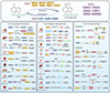 |
Figure 1 The main components of m6A modification and the mechanism of the corresponding molecules in tumor autophagy and apoptosis, metabolic reprogramming and tumor microenvironment. This figure created by Figdraw (https://www.figdraw.com/). |
Architectural framework of m6A regulatory machinery
The discovery of fat mass and obesity-associated protein (FTO) as the first m6A demethylase in 2011 revolutionized our understanding of RNA epigenetics by establishing m6A as a reversible modification [19]. This breakthrough was followed by the identification of RRACH (R=G/A; H=A/C/U) as the consensus motif, predominantly localized in 3’UTRs and final exons of non-coding RNAs through high-throughput sequencing [2, 20]. The core regulatory mechanism of m6A modification involves coordinated interactions among three functional protein groups: “Writers”, “Erasers”, and “Readers”, which collectively mediate the dynamic deposition, removal, and interpretation of this RNA modification [21].
m6A writers
The installation of m6A modifications is catalyzed by a methyltransferase complex composed of Methyltransferase-like 3 (METTL3), Methyltransferase-like 14 (METTL14), and Wilms’ tumor 1-associating protein (WTAP) [22]. METTL3 is the SAM-binding catalytic core for m6A deposition, while METTL14 stabilizes the heterodimer through its RGG domain and enhances RNA binding, forming a composite active site that targets specific transcripts. On one hand, this complex can control the stability of target mRNAs through an m6A-dependent mechanism, i.e., recognition by reader YTH domain-containing family protein [23]. Meanwhile, it can also promote oncogene expression through an m6A-independent manner, in which cytoplasmic METTL3 directly recruits translation machinery (e.g., eukaryotic initiation factor 3, eIF3) [24]. The abnormal upregulation of METTL3/14 in cancer alters the m6A landscape on key mRNAs (MYC, MYB, and PES1), thereby driving oncogene translation and leukemogenesis [25]. WTAP stabilizes this core complex and facilitates its localization to nuclear speckles [26]. Additionally, RNA-binding motif protein 15 (RBM15), Vir-like m6A methyltransferase-associated protein (VIRMA), and Zinc finger CCCH-type containing 13 (ZC3H13) are recognized as components of the m6A methyltransferase complex [27], though only METTL3 exhibits catalytic activity [28].
m6A erasers
The RNA m6A demethylases – fat mass and obesity-associated protein (FTO), ALKB homolog 5 (ALKBH5), and ALKB homolog 3 (ALKBH3) – belong to the ALKB dioxygenase family and require α-ketoglutarate and Fe2⁺ as cofactors to catalyze m6A removal [29]. Structurally, these enzymes feature shallow substrate-binding clefts, restricting their activity primarily to single-stranded RNAs such as mRNA [30]. FTO dynamically regulates RNA metabolism (splicing, stabilization, and translation) mainly by demethylating the m6A modification inside the mRNA and has secondary activity on the cap m6Am under specific circumstances [31]. Current research confirms that ALKBH5 mediates m6A demethylation in mRNAs and lncRNAs/non-coding RNAs [32, 33], while ALKBH3, the newest m6A demethylase identified [34], is also involved in N1-methyladenine (m1A) RNA modification pathways [35]. The dynamic reversibility of m6A modifications is governed by the interplay between “writers” and “erasers”, while their biological functions ultimately depend on recognition by “readers” [36].
m6A readers
The YT521-B homology (YTH) domain-containing proteins represent a fundamental class of m6A reader proteins that play pivotal roles in RNA metabolism regulation [37–39]. Among these, YTH domain-containing family protein 2 (YTHDF2) specifically recognizes m6A-modified RNA through its characteristic C-terminal aromatic cage structure, composed of conserved residues including Trp432 and Trp486 [40]. This recognition enables YTHDF2 to facilitate RNA degradation through two distinct mechanisms: either by direct recruitment of the CCR4/NOT deadenylase complex via its N-terminal domain, or through the YTHDF2-HRSP12-RNase P/MRP pathway [41]. In contrast, YTHDF1 employs a similar aromatic cage structure (containing Trp411) for m6A recognition but functions primarily in translation activation through its interaction with eukaryotic initiation factor 3 (eIF3) [42]. YTHDF3 cooperates with both YTHDF1 and YTHDF2 to regulate RNA translation or degradation [43, 44]. The activities of these YTH domain proteins are subject to sophisticated cellular regulation: YTHDF2 stability is enhanced through ERK1/2-mediated phosphorylation under hypoxic conditions, while its transcriptional expression is modulated by histone H3K18 lactylation [45]. Furthermore, competitive interactions between different m6A readers, such as the competition between YTHDF proteins and Insulin-like growth factor 2 mRNA-binding proteins (IGF2BPs) for m6A binding sites on c-Myc mRNA, create an additional layer of regulatory complexity in determining RNA processing outcomes [46].
Other YTH family members include YTH domain-containing 1 (YTHDC1) and YTHDC2, which mediate RNA splicing or degradation through m6A recognition [47]. Beyond YTH domain proteins, additional proteins serve as m6A readers. For instance, IGF2BPs (IGF2BP1/2/3) recognize m6A modifications primarily within the 3’ UTRs of target mRNAs, where they recruit stabilizing factors and translation enhancers to suppress deadenylation and promote polysome assembly, thereby enhancing mRNA stability and translation efficiency [48]. Heterogeneous nuclear ribonucleoproteins (HNRNPs) such as HNRNPA2/B1 facilitate the processing of primary microRNA (pri-miRNA) transcripts by recognizing m6A marks [49], while HNRNPC and HNRNPG regulate mRNA abundance and splicing through m6A-dependent mechanisms [50–52]. Nevertheless, the precise molecular mechanisms by which these factors coordinate m6A-mediated regulation of cellular processes and disease pathogenesis remain to be fully elucidated.
The regulatory role of m6A modifications in tumor autophagy and apoptosis
Tumor autophagy
Autophagy represents an evolutionarily conserved catabolic process essential for eukaryotic cellular homeostasis [53]. Dysregulated autophagy is closely associated with diverse physiological and pathological processes, including cellular homeostasis, aging, neurodegenerative disorders, and cancer [54]. Autophagy is broadly categorized into three types – macroautophagy, microautophagy, and chaperone-mediated autophagy – all operating through the autophagic-lysosomal pathway (ALP) [55, 56]. Among these, macroautophagy (hereafter referred to as autophagy) represents the predominant form [55]. Studies demonstrate that during early tumorigenesis, autophagy exerts tumor-suppressive effects through its dual functionality as a cell survival pathway and a quality control mechanism, effectively inhibiting tumor initiation and progression [57]. However, when tumors progress to advanced stages and establish under environmental stress, autophagy undergoes functional reprogramming into a dynamic degradation and recycling system. This transition not only sustains the survival and proliferation of primary tumors but also promotes aggressive cancer phenotypes by enhancing metastatic potential [57].
The autophagic process is primarily divided into four distinct phases: initiation, formation of the isolation membrane and autophagosome, autophagosome-lysosome fusion, and autophagic degradation [58], with each phase being regulated by epigenetic modifications [59]. Among these regulatory mechanisms, m6A modification plays a pivotal role in modulating tumor-associated autophagy [60]. The mechanistic target of rapamycin (mTOR), a key regulator of autophagy initiation, serves as a critical downstream effector of the Phosphatidylinositol 3-kinase (PI3K)/AKT (Protein Kinase B) signaling pathway [61]. In endometrial cancer, m6A modification has been shown to regulate AKT kinase activity, suggesting its involvement in autophagy modulation through the AKT-mTOR signaling axis [62]. Subsequent studies have further established the role of m6A in autophagy regulation across various malignancies.
For instance, in non-small cell lung cancer (NSCLC), upregulation of the m6A demethylase ALKBH5 drives tumor progression by reducing m6A levels on UBE2C mRNA, thereby disrupting the UBE2C-mediated autophagy suppression axis [63]. In small cell lung cancer, the m6A methyltransferase METTL3 promotes chemoresistance by targeting DCP2 to regulate Pink1-Parkin pathway-mediated mitophagy and mitochondrial dysfunction [64]. A study about hepatocellular carcinoma (HCC) reveals that METTL3 modulates sorafenib resistance through m6A-dependent regulation of autophagy transcription factor FOXO3 under hypoxic tumor microenvironments [65]. Hypoxia-inducible factor 1α (HIF-1α) further enhances hepatocellular carcinogenesis by inducing YTHDF1 expression, which drives autophagy and cancer progression via translational upregulation of ATG2A and ATG14 [66]. In bladder cancer, METTL16 binds to m6A sites in the 3’-UTR of PMEPA1 mRNA, reducing its stability to suppress tumor proliferation while enhancing cisplatin sensitivity through PMEPA1-mediated autophagy pathways [67]. In colon cancer, autophagy caused by insufficient cell energy promotes the expression of m6A reading protein IGF2BP2, stabilizes MYC mRNA, and then promotes the glycolysis process of tumor cells, thereby increasing cell energy supply and promoting the proliferation of cancer cells [68]. Melanoma studies highlight the critical role of the m6A demethylase FTO in therapeutic resistance, where nutrient-deprived tumor microenvironments induce autophagy-mediated FTO upregulation. This reduces m6A levels on PD-1, CXCR4, and SOX10 mRNAs, attenuating YTHDF2-mediated degradation and potentiating immune evasion [69].
In summary, accumulating evidence demonstrates that m6A RNA methylation participates in dysregulated autophagy signaling and plays a critical role in the pathogenesis and progression of diverse malignancies. Therefore, comprehensive elucidation of the regulatory interplay between m6A modifications and autophagy-related molecules, as well as their functional implications in cancer advancement and therapeutic responses, will not only advance our understanding of novel oncogenic mechanisms but also provide a critical theoretical framework for developing targeted therapeutic strategies centered on the m6A-autophagy regulatory axis.
Tumor apoptosis
Apoptosis, a canonical form of programmed cell death, plays a fundamental role in maintaining tissue homeostasis by eliminating harmful cells with DNA damage or aberrant proliferation [70]. In recent years, the critical involvement of apoptotic regulation in tumorigenesis and cancer progression has garnered substantial research attention. Emerging evidence indicates that neoplastic cells acquire apoptosis resistance mechanisms to evade immune surveillance and programmed cell death, conferring not only significant survival advantages but also promoting malignant transformation and disease progression. Notably, such apoptotic resistance represents a pivotal factor contributing to the failure of conventional chemotherapy and targeted therapies, posing a major scientific challenge in current oncology therapeutics [71].
Recent studies have revealed the crucial regulatory role of m6A modification in tumor cell apoptosis, though mechanistic investigations remain relatively limited. The m6A “writers”, “erasers”, and “readers” orchestrate apoptosis through context-dependent mechanisms across cancers.
The m6A methyltransferase METTL3 has been demonstrated to be intimately associated with the initiation and maintenance of acute myeloid leukemia (AML). Strikingly, its specific inhibitor STM2457 markedly suppresses leukemic cell proliferation while inducing apoptosis and differentiation [72]. Similarly, loss of METTL3 in NSCLC enhances cisplatin-induced apoptosis by upregulating BAX and PUMA gene via reduced m6A methylation on their transcripts [73]. A recent study shows that the stapled peptide inhibitor RSM3 targeting METTL3-METTL14 binding inhibits complex formation, promotes METTL3 proteasomal degradation, reduces global RNA methylation, upregulates apoptosis-related genes, suppresses pro-cancer signals, and inhibits tumor growth with enhanced apoptosis in vivo, offering a novel mechanism distinct from small-molecule inhibitors for METTL3-targeted cancer therapy [74].
Among m6A demethylases, decreased ALKBH5 expression correlates with poor prognosis in osteosarcoma patients. Functional studies show that ALKBH5 upregulation reduces m6A levels in osteosarcoma cells, consequently inhibiting proliferation and promoting apoptosis alongside cell cycle arrest [75]. In pancreatic cancer, ALKBH5 loss drives cancer progression via YTHDF2-dependent PER1 m6A demethylation, inhibiting apoptosis through the ATM-CHK2-P53 pathway [76]. Conversely, FTO mediates fluorouracil (5-FU) resistance in colorectal cancer through m6A-dependent regulation of the apoptotic gene SIVA1 [77]. Notably, in prostate cancer, IGF2BP1 stabilizes NSMCE2 mRNA in an m6A-dependent manner by recognizing VIRMA-mediated methylation, thereby reducing reactive oxygen species (ROS) levels and apoptosis to promote tumor progression [78].
In all, the dynamic interplay between m6A RNA methylation, autophagy, and apoptosis highlights its critical role in tumor biology as a key link for cancer progression and treatment resistance. Emerging evidence highlights that m6A-modifying enzymes orchestrate context-dependent regulatory networks that influence autophagy and apoptosis, with important effects on tumor survival, metastasis, and drug sensitivity. However, substantial challenges persist, encompassing the dual roles of m6A regulators in mediating autophagy-apoptosis crosstalk and the tissue-specific heterogeneity of their functional outcomes. Future investigative priorities should focus on deciphering the spatiotemporal kinetics of m6A regulation, its synergistic or antagonistic interactions with other epigenetic modalities, and the rational design of therapeutic interventions tailored to specific tumor microenvironments. Targeting the m6A-autophagy-apoptosis axis holds significant translational potential for overcoming therapeutic resistance, yet its clinical realization hinges on resolving the paradoxical functions of these enzymes in malignant transformation and microenvironmental modulation.
The role of m6A modification in tumor metabolic reprogramming
Early studies have established cancer as a genetic disease, with nearly all cancers driven by genetic alterations. Substantial evidence indicates that all cancer cells exhibit six core biological capabilities: sustaining proliferative signaling, evading growth suppressors, resisting cell death and apoptosis, achieving limitless replicative potential, inducing angiogenesis, and promoting invasion and metastasis [79]. Subsequent research has identified two additional canonical traits-metabolic reprogramming and immune evasion [71]. The latest advances further expand this conceptual framework by introducing new hallmarks and enabling characteristics, including phenotypic plasticity, nonmutational epigenetic reprogramming, polymorphic microbiomes, and the functional role of senescent cells in the tumor microenvironment [80]. Accumulating evidence indicates that m6A modifications participate in modulating key metabolic pathways in malignancies, including glucose, fatty acid, and amino acid metabolism. Consequently, cancer can no longer be viewed solely through a genetic lens but must also be understood as a metabolic disease. Exploring the mechanism of metabolic reprogramming of cancer is helpful for a better understanding of the oncogenesis and progression process, thereby facilitating the identification of effective therapeutic targets for cancer.
Glucose metabolism
As the most extensively studied facet of tumor metabolic reprogramming, glucose metabolism exemplifies how m6A modification reshapes cancer cell bioenergetics. Dysregulated glucose metabolism, a hallmark of tumor metabolic reprogramming, manifests as enhanced glycolytic flux and lactate fermentation (the Warburg effect). This metabolic adaptation empowers cancer cells to mitigate metabolic stress, thrive in hostile microenvironments, and sustain malignant proliferation [81]. Mechanistically, m6A regulators orchestrate glucose metabolism through both upstream signaling and direct modulation of glycolytic effectors. Investigations in temozolomide-resistant gliomas reveal that the Warburg effect promotes extracellular vesicle-mediated release of circ-0072083, which targets the miR-1252-5p/ALKBH5 axis to reduce m6A modification on NANOG mRNA, a core pluripotency transcription factor, thereby stabilizing its expression and exacerbating chemoresistance [82]. In colorectal cancer, overexpression of the m6A reader IGF2BP2 stabilizes the ZFAS1/OLA1 axis, augmenting obg-like ATPase 1 (OLA1) recruitment, ATP hydrolysis, and glycolytic activity to activate the Warburg effect, ultimately enhancing tumor cell proliferation and clonogenicity [83]. Furthermore, in NSCLC, METTL3-mediated m6A methylation of the long non-coding RNA ABHD11-AS1 enhances its stability and expression, consequently stimulating Warburg metabolism and malignant cell growth [84].
At the effector level, m6A directly fine-tunes rate-limiting enzymes and transporters to drive glycolytic dependency. For instance, METTL3-mediated m6A modification at the 3’UTR of hexokinase 2 (HK2) mRNA, the rate-limiting enzyme in glycolysis, cooperates with YTHDF1 to stabilize its transcript, thereby potentiating the Warburg effect in cervical carcinogenesis [85]. METTL3 further orchestrates m6A-dependent regulation of the glucose transporter isoform 1 (SLC2A1; GLUT1), relying on IGF2BP2/3 to stabilize its expression and activate downstream glycolytic pathways, ultimately driving colorectal cancer progression [86]. In bladder cancer, ALKBH5 downregulation modulates m6A-dependent stability of casein kinase 2 alpha subunit (CK2α) mRNA, suppressing CK2α-mediated glucose uptake, lactate/ATP production, and tumorigenesis while enhancing cisplatin sensitivity [87]. Additionally, key metabolic regulators, including pyruvate kinase M2 (PKM2), cellular myelocytomatosis oncogene (C-Myc), and non-canonical glycolytic enzymes like pyruvate dehydrogenase kinase 1 (PDK1), are subject to m6A-mediated regulation, collectively shaping tumor progression trajectories [88–90].
Lipid metabolism
Within the tumor microenvironment, nutrient deprivation drives cancer cells to reprogram lipid metabolism to sustain proliferation, survival, invasion, and metastatic capabilities [91]. Beyond glucose metabolism, m6A modification emerges as a key orchestrator of lipid metabolic reprogramming, enabling cancer cells to survive nutrient-deprived microenvironments. Studies demonstrate that FTO knockout significantly reduces de novo lipogenesis enzyme levels and lipid content in HCC cells. Mechanistically, FTO ablation elevates m6A modification on fatty acid synthase (FASN) mRNA, which is recognized by YTHDF2 to destabilize the transcript, thereby reducing protein levels of acetyl-CoA carboxylase (ACC) and ATP-citrate lyase (ACLY). This lipid biosynthesis impairment ultimately induces HCC cell death due to insufficient lipid accumulation [92].
In colorectal cancer, YTHDF1 enhances the mRNA stability of DDX21 by recognizing its m6A modification site, which further binds to the promoter region of 3-hydroxy-3-methylglutaryl-coa synthase 1 (HMGCS1) and directly activates HMGCS1 transcription to promote tumor progression [93]. Furthermore, in esophageal squamous cell carcinoma (ESCC), the m6A reader HNRNPA2B1 upregulates fatty acid metabolism-related genes ACLY and ACC1, facilitating lipid droplet formation and promoting tumor cell proliferation, migration, and invasion [94]. In ovarian cancer, the m6A reading protein IGF2BP3 promotes cholesterol synthesis and inhibits lipolysis by regulating the ESM1/KLF10/BECN1 feedback loop, thereby promoting tumor progression [95]. These findings collectively illustrate how m6A readers establish a pro-lipogenic state to support tumor cell bioenergetics and membrane synthesis.
Amino acid metabolism
The m6A modification also intersects with amino acid metabolism, linking nutrient availability to epigenetic reprogramming and metastatic potential. In clear cell renal cell carcinoma, the m6A RNA demethylase FTO forms a synthetic lethal partnership with VHL through m6A modification-dependent regulation of the glutamine transporter SLC1A5. FTO-mediated demethylation of SLC1A5 mRNA enhances its stability and expression, thereby promoting glutamine uptake and metabolic reprogramming in VHL-deficient cells [96]. Similarly, ALKBH1 functions as an m6A demethylase, catalyzing demethylation at critical residues (Y184, H231, D233) to enhance lung cancer cell migration and invasion [97]. Notably, this regulatory axis extends to branched-chain amino acid metabolism.
Branched-chain amino acids catabolism depletes α-KG levels, suppressing ALKBH5 demethylase function and consequently elevating m6A methylation on EMT-related transcripts, ultimately promoting NSCLC brain metastasis through this epigenetic pathway [98]. Similarly, the curcumin analog WZ35 disrupts glutamine metabolism by targeting the GLS2-ALKBH5-m6A regulatory axis, leading to glutathione depletion and consequent suppression of gastric cancer metastasis [99]. In AML, the m6A reader protein IGF2BP2 sustains leukemia stem cells through m6A-dependent regulation of glutamine metabolism genes (MYC, GPT2, SLC1A5). Pharmacological targeting of IGF2BP2 with CWI1-2 disrupts this metabolic axis, thereby offering a promising therapeutic strategy for AML [100]. These studies reveal a bidirectional relationship between amino acid availability, m6A dynamics, and tumor cell plasticity.
The role of m6A modification in the tumor microenvironment
Immunosuppression constitutes a central hallmark of the tumor microenvironment (TME), primarily characterized by functional exhaustion of cytotoxic immune cells, dysfunction of antigen-presenting cells (APCs), and extensive recruitment or induction of immunosuppressive cell populations. These inhibitory immune components include CD4+ regulatory T cells (Tregs), immature myeloid cells (iMCs)/myeloid-derived suppressor cells (MDSCs), tumor-associated macrophages (TAMs), and various immunosuppressive cytokines [101, 102]. In addition, endothelial cells in the tumor microenvironment mediate tumor immunosuppression by expressing immunosuppressive molecules [103], inhibiting leukocyte adhesion/activation, and recruiting immunosuppressive cells [104], which ultimately promote immune escape and tumor progression [105]. During tumor progression, infiltrating immune cells undergo remodeling processes that profoundly impact their intrinsic biological functions. These alterations can facilitate immune evasion while sustaining the proliferative capacity of tumor cells [102]. Emerging evidence suggests that m6A modification may be implicated in tumor immune remodeling processes through multifaceted regulatory mechanisms.
Regulation of immune cell function
The m6A machinery differentially modulates immune cell activity through distinct molecular pathways. For instance, YTHDF2 binds m6A-modified STAT1 mRNA in TAMs, promoting its decay. Inhibition of YTHDF2 activates IFNγ-STAT1 signaling to reprogram TAMs, thereby enhancing CD8+ T cell-mediated antitumor immunity and offering a potential strategy to improve immune checkpoint therapy efficacy [106]. METTL3-mediated m6A modification enables YTHDF1 to promote SPRED2 translation, suppressing NF-κB/STAT3 pathways in TAMs [107]. Furthermore, YTHDF1 modulates dendritic cells (DCs) by regulating the translation of lysosomal proteases via m6A modification, impairing tumor antigen cross-presentation and CD8+ T cell cross-priming, thereby limiting antitumor immunity [108].
Tumor-intrinsic immune modulation
In addition to immune cells, intrinsic m6A modifications in tumor cells profoundly influence immune remodeling processes in the TME. Studies reveal that inhibition of m6A methyltransferases METTL3 and METTL4 stabilizes STAT1 and IRF1 mRNAs through a YTHDF2-dependent mechanism, activating the IFN-γ-STAT1-IRF1 signaling axis. This activation enhances CD8+ T cell infiltration and cytokine secretion, thereby potentiating the efficacy of PD-1 blockade therapy [109]. In melanoma, FTO-mediated m6A demethylation contributes to immunotherapy resistance by upregulating key genes, including PD-1, CXCR4, and SOX10. Conversely, pharmacological inhibition of FTO enhances tumor responsiveness to IFN-γ and overcomes PD-1 inhibitor resistance [69]. Furthermore, YTHDF1 facilitates melanoma immune evasion and resistance to immune checkpoint inhibitors by promoting degradation of major histocompatibility complex class I (MHC-I) molecules [110].
In colon cancer, YTHDF1 enhances p65 translation via m6A modification, subsequently elevating CXCL1 expression. This upregulation recruits MDSCs through the CXCL1-CXCR2 axis, thereby accelerating tumor progression and immunotherapy resistance [17]. Breast cancer studies demonstrate that the METTL3/IGF2BP3 axis upregulates PD-L1 expression via m6A modification, enabling cancer cell immune evasion [111]. In bladder cancer, JNK signaling activates c-Jun to promote METTL3 expression, which stabilizes PD-L1 mRNA through m6A modification, amplifying PD-L1 expression and fostering immune escape. Inhibition of the JNK/METTL3 axis reduces PD-L1 levels and enhances CD8+ T cell-mediated antitumor activity [112]. Additionally, loss or inhibition of the m6A demethylase ALKBH5 enhances tumor sensitivity to immune checkpoint blockade (ICB) therapy by remodeling lactate metabolism and reshaping immunosuppressive cellular components within the TME, thereby potentiating immunotherapeutic outcomes [113]. Targeting the demethylase FTO has also been found to suppress cancer stem cell properties and immune evasion mechanisms [114]. These findings collectively unveil a multidimensional regulatory network of m6A modifications in tumor cell-TME interactions.
Vascular remodeling
Accumulating evidence highlights the pivotal role of m6A RNA modification in modulating endothelial cell plasticity and tumor vascularization. In hypoxic breast cancer microenvironments, RPPH1 upregulation stabilizes FGFR2 mRNA via the IGF2BP2-m6A axis, activating PI3K/AKT signaling to enhance EC-driven angiogenesis, cancer stemness, and metastasis [115]. Similarly, IGF2BP2 interacts with FLT4 in lung adenocarcinoma, amplifying PI3K/AKT phosphorylation to promote EC proliferation and vascular lumen formation [116]. Beyond mRNA stabilization, the m6A reader YTHDF3 facilitates breast cancer brain metastasis by enhancing the translation of m6A-enriched transcripts (e.g., ST6GALNAC5, GJA1, and EGFR), thereby strengthening tumor cell adhesion to brain ECs and promoting transendothelial invasion [117]. Additionally, METTL3-mediated m6A deposition coordinates IGF2BP2/3-dependent stabilization of EphA2 and VEGFA in colorectal cancer, activating PI3K/AKT/mTOR and ERK1/2 pathways to drive vasculogenic mimicry (VM), a process enabling tumor cells to adopt EC-like phenotypes and form perfusion-supporting channels [118]. Collectively, these findings highlight m6A machinery as a multifaceted regulator of EC-tumor crosstalk, driving angiogenesis, metastatic niche formation, and therapy-resistant vascular remodeling, thus presenting novel targets for anti-cancer strategies.
Conclusion and future perspectives
In recent years, m6A modification, as a central research focus in epitranscriptomics, has increasingly been recognized for its regulatory roles in tumorigenesis. This review systematically delineates the dynamic regulatory mechanisms of m6A modification and its multifaceted roles in tumor progression. First, it outlines the molecular basis of m6A modification, which involves three core enzyme classes, writers (METTL3/14/16, WTAP, VRIMA, RBM15, ZC3H13), erasers (FTO, ALKBH3/5), and readers (YTHDFs, YTHDCs, IGF2BPs, HNRNPA2B1, MSI2), that dynamically regulate RNA stability, translation efficiency, and subcellular localization, thereby influencing tumor cell proliferation, metastasis, and drug resistance. Subsequently, this review comprehensively discusses the regulatory roles of m6A modification in tumor autophagy and apoptosis, metabolic reprogramming, and tumor microenvironment dynamics.
This review reveals that m6A-modifying enzymes (writers, erasers, and readers) exhibit paradoxical tumor-regulatory roles across cancer types and microenvironments. For example, METTL3 promotes sorafenib resistance via FOXO3-mediated autophagy in hypoxic HCC [65], but enhances cisplatin sensitivity by upregulating BAX/PUMA in NSCLC [73]. ALKBH5 drives NSCLC progression by activating UBE2C-mediated autophagy [63], yet suppresses osteosarcoma growth via global m6A reduction [75]. FTO enables melanoma immune evasion by stabilizing PD-1/CXCR4 mRNAs [69], while inducing HCC cell death via FASN-dependent lipid biosynthesis impairment [92]. These contradictions highlight context-dependent targeting of survival/apoptotic, metabolic/immune pathways, underscoring the complexity of epitranscriptomic regulation in cancer.
Although significant progress has been made in understanding m6A modifications in oncology, several critical gaps and challenges remain. Firstly, emerging evidence suggests that m6A-modifying enzymes themselves are epigenetically regulated, which is a complex interaction that requires systematic study. Moreover, the substantial heterogeneity among tumors necessitates further elucidation of tumor-type-specific m6A regulatory mechanisms. Additionally, recent studies have uncovered non-canonical m6A regulators, such as non-canonical cap-binding protein eIF3d, mediating stress response via ALKBH5-dependent demethylation [119], IGF2BP1 recognizing m6A through a hydrophobic platform independent of YTH proteins [120], METTL14 binding to RNA G-quadruplexes to guide methylation [121], and the RNA helicase DDX21 directs co-transcriptional m6A deposition by interacting with METTL3 to recognize R-loops and promote RNAPII termination [122], highlighting emerging regulatory mechanisms beyond canonical writer/reader/eraser pathways.
From a translational standpoint, four strategic priorities emerge: (1) Systematic identification of m6A regulators and their downstream targets as dual-function biomarkers for both prognostic evaluation and immunotherapy response prediction; (2) Development of precision therapeutics combining m6A-targeted small-molecule modulators with immune checkpoint blockade therapies to achieve synergistic antitumor efficacy; (3) Establishment of dynamic m6A regulatory network models through artificial intelligence-powered multi-omics integration, enabling comprehensive analysis of tumor progression mechanisms; (4) Implementing clinically representative model systems, particularly patient-derived xenograft (PDX) models and patient-derived organoid (PDO) models, combined with multi-omics validation using clinical specimens. This integrated approach will facilitate the translation of fundamental discoveries into clinical applications, ultimately advancing precision therapeutic strategies for cancer patients.
In conclusion, as a critical bridge linking the epitranscriptome with functional tumor phenotypes, m6A modification research has not only deepened our understanding of malignant biological behaviors in oncology but also established theoretical frameworks for developing novel diagnostic tools and combination therapeutic strategies. Future investigations should prioritize two synergistic dimensions: mechanistic depth and clinical translation.
Funding
This work was supported by the National Natural Science Foundation of China (No. 82172716), the China Society of Clinical Oncology Foundation (No. 2022143, No. 2024218), and the Wu Jieping Medical Foundation (No. 2022140).
Conflicts of interest
The authors declare no conflict of interest.
Data availability statement
This review has no associated data generated.
Author contribution statement
Conceptualization, S.X., Y.L.; Writing-original draft, Y.L., K.Z.; Writing-reviewing, Y.F., S.Y., Y.H., X.X., Z.H.; Writing-reviewing and editing, Y.L., K.Z., S.X.
Ethics approval
Ethical approval was not required.
References
- Liu N, Pan T. N6-methyladenosine-encoded epitranscriptomics. Nature Structural and Molecular Biology. 2016;23(2):98–102. [Google Scholar]
- Meyer KD, Saletore Y, Zumbo P, et al. Comprehensive analysis of mRNA methylation reveals enrichment in 3’ UTRs and near stop codons. Cell. 2012;149(7):1635–1646. [Google Scholar]
- Wang X, Zhao BS, Roundtree IA, et al. N(6)-methyladenosine modulates messenger rna translation efficiency. Cell. 2015;161(6):1388–1399. [Google Scholar]
- Wang T, Kong S, Tao M, et al. The potential role of RNA N6-methyladenosine in Cancer progression. Molecular Cancer. 2020;19(1): 88. [Google Scholar]
- Zhang Z, Zhou D, Qiu X, et al. N6-methyladenosine-mediated EIF3H promotes anaplastic thyroid cancer progression and ferroptosis resistance by stabilizing β-catenin. Free Radical Biology and Medicine. 2025;231:38–47. [Google Scholar]
- Wang J, Yu H, Dong W, et al. N6-methyladenosine-mediated up-regulation of FZD10 regulates liver cancer stem cells’ properties and Lenvatinib resistance through WNT/β-catenin and hippo signaling pathways. Gastroenterology. 2023;164(6):990–1005. [Google Scholar]
- Chen X, Wang J, Tahir M, et al. Current insights into the implications of m6A RNA methylation and autophagy interaction in human diseases. Cell and Bioscience. 2021;11(1):147. [Google Scholar]
- Liu L, Li H, Hu D, et al. Insights into N6-methyladenosine and programmed cell death in cancer. Molecular Cancer. 2022;21(1):32. [Google Scholar]
- An Y, Duan H. The role of m6A RNA methylation in cancer metabolism. Molecular Cancer. 2022;21(1):14. [Google Scholar]
- Li M, Zha X, Wang S. The role of N6-methyladenosine mRNA in the tumor microenvironment. Biochimica et Biophysica Acta. Reviews on Cancer. 2021;875(2):188522. [Google Scholar]
- Zhuang H, Yu B, Tao D, et al. The role of m6A methylation in therapy resistance in cancer. Molecular Cancer. 2023;22(1):91. [Google Scholar]
- Li Y, Guo M, Qiu Y, et al. Autophagy activation is required for N6-methyladenosine modification to regulate ferroptosis in hepatocellular carcinoma. Redox biology. 2024;69:102971. [Google Scholar]
- Lai X, Wei J, Gu X, et al. Dysregulation of LINC00470 and METTL3 promotes chemoresistance and suppresses autophagy of chronic myelocytic leukaemia cells. Journal of Cellular and Molecular Medicine. 2021;25(9):4248–4259. [Google Scholar]
- Yue S, Liu H, Su H, et al. m6A-regulated tumor glycolysis: new advances in epigenetics and metabolism. Molecular Cancer. 2023;22(1):137. [Google Scholar]
- Yang Y, Cai J, Yang X, et al. Dysregulated m6A modification promotes lipogenesis and development of non-alcoholic fatty liver disease and hepatocellular carcinoma. Molecular Therapy. 2022;30(6):2342–2353. [Google Scholar]
- Dai C, Cao J, Tang Y, et al. YTHDF3 phase separation regulates HSPA13-dependent clear cell renal cell carcinoma development and immune evasion. Cancer Science. 2024;115(8):2588–2601. [Google Scholar]
- Bao Y, Zhai J, Chen H, et al. Targeting m(6)A reader YTHDF1 augments antitumour immunity and boosts anti-PD-1 efficacy in colorectal cancer. Gut. 2023;72(8):1497–1509. [Google Scholar]
- Shriwas O, Mohapatra P, Mohanty S, et al. The impact of m6A RNA modification in therapy resistance of cancer: implication in chemotherapy, radiotherapy, and immunotherapy. Frontiers in Oncology. 2020;10:612337. [Google Scholar]
- Jia G, Fu Y, Zhao X, et al. N6-methyladenosine in nuclear RNA is a major substrate of the obesity-associated FTO. Nature Chemical Biology. 2011;7(12):885–887. [Google Scholar]
- Dominissini D, Moshitch-Moshkovitz S, Schwartz S, et al. Topology of the human and mouse m6A RNA methylomes revealed by m6A-seq. Nature. 2012;485(7397):201–206. [Google Scholar]
- He L, Li H, Wu A, et al. Functions of N6-methyladenosine and its role in cancer. Molecular Cancer. 2019;18(1):176. [Google Scholar]
- Liu J, Yue Y, Han D, et al. A METTL3-METTL14 complex mediates mammalian nuclear RNA N6-adenosine methylation. Nature Chemical Biology. 2014;10(2):93–95. [Google Scholar]
- Wang X, Feng J, Xue Y, et al. Structural basis of N(6)-adenosine methylation by the METTL3-METTL14 complex. Nature. 2016;534(7608):575–578. [Google Scholar]
- Choe J, Lin S, Zhang W, et al. mRNA circularization by METTL3-eIF3h enhances translation and promotes oncogenesis. Nature. 2018;561(7724):556–560. [Google Scholar]
- Du W, Huang Y, Chen X, et al. Discovery of a PROTAC degrader for METTL3-METTL14 complex. Cell Chemical Biology. 2024;31(1):177–183. [Google Scholar]
- Ping X, Sun B, Wang L, et al. Mammalian WTAP is a regulatory subunit of the RNA N6-methyladenosine methyltransferase. Cell Research. 2014;24(2):177–189. [Google Scholar]
- Jiang X, Liu B, Nie Z, et al. The role of m6A modification in the biological functions and diseases. Signal Transduction and Targeted Therapy. 2021;6(1):74. [Google Scholar]
- Wang X, Huang J, Zou T, et al. Human m(6)A writers: two subunits, 2 roles. RNA Biology. 2017;14(3):300–304. [Google Scholar]
- Zheng G, Dahl JA, Niu Y, et al. ALKBH5 is a mammalian RNA demethylase that impacts RNA metabolism and mouse fertility. Molecular Cell. 2013;49(1):18–29. [Google Scholar]
- Xu B, Liu D, Wang Z, et al. Multi-substrate selectivity based on key loops and non-homologous domains: new insight into ALKBH family. Cellular and Molecular Life Sciences. 2021;78(1): 129–141. [Google Scholar]
- Yen YP, Lung TH, Liau ES, et al. The motor neuron m6A repertoire governs neuronal homeostasis and FTO inhibition mitigates ALS symptom manifestation. Nature Communications. 2025;16(1):4063. [Google Scholar]
- Qu S, Feng B, Xing M, et al. PRMT5 K240lac confers ferroptosis resistance via ALKBH5/SLC7A11 axis in colorectal cancer. Oncogene. 2025. https://doi.org/10.1038/s41388-025-03457-2. [Google Scholar]
- Chen Z, Zhou Y, Xue C, et al. Psychological stress-induced ALKBH5 deficiency promotes tumour innervation and pancreatic cancer via extracellular vesicle transfer of RNA. Nature Cell Biology. 2025;27:1035–1047. [Google Scholar]
- Ueda Y, Ooshio I, Fusamae Y, et al. AlkB homolog 3-mediated tRNA demethylation promotes protein synthesis in cancer cells. Scientific Reports. 2017;7:42271. [Google Scholar]
- Tu L, Gu S, Xu R, et al. ALKBH3-mediated M1A demethylation of METTL3 endows pathological fibrosis: interplay between M1A and M6A RNA methylation. Advanced Science (Weinheim, Baden-Württemberg, Germany). 2025;12(19):e2417067. [Google Scholar]
- Wang X, Lu Z, Gomez A, et al. N6-methyladenosine-dependent regulation of messenger RNA stability. Nature. 2014;505(7481):117–120. [Google Scholar]
- Liao J, Wei Y, Liang J, et al. Insight into the structure, physiological function, and role in cancer of m6A readers-YTH domain-containing proteins. Cell Death Discovery. 2022;8(1):137. [Google Scholar]
- Yang H, Chiang C, Luo Q, et al. YT521-B homology domain family proteins as N6-methyladenosine readers in tumors. Frontiers in Genetics. 2022;13:934223. [Google Scholar]
- Wang J, Lu A. The biological function of m6A reader YTHDF2 and its role in human disease. Cancer Cell International. 2021;21(1):109. [Google Scholar]
- Du H, Zhao Y, He J, et al. YTHDF2 destabilizes m(6)A-containing RNA through direct recruitment of the CCR4-NOT deadenylase complex. Nature Communications. 2016;7:12626. [Google Scholar]
- Temme C, Zhang L, Kremmer E, et al. Subunits of the Drosophila CCR4-NOT complex and their roles in mRNA deadenylation. RNA. 2010;16(7):1356–1370. [Google Scholar]
- Zaccara S, Jaffrey SR. A unified model for the function of YTHDF proteins in regulating m6A-modified mRNA. Cell. 2020;181(7): 1582–1595.e18. [Google Scholar]
- Shi H, Wang X, Lu Z, et al. YTHDF3 facilitates translation and decay of N(6)-methyladenosine-modified RNA. Cell Research. 2017;27(3):315–328. [Google Scholar]
- Zhao Y, Huang S, Tan X, et al. N(6)-Methyladenosine-modified CBX1 regulates nasopharyngeal carcinoma progression through heterochromatin formation and STAT1 activation. Advanced Science. 2022;9(36):e2205091. [Google Scholar]
- Xu P, Hu K, Zhang P, et al. Hypoxia-mediated YTHDF2 overexpression promotes lung squamous cell carcinoma progression by activation of the mTOR/AKT axis. Cancer Cell Int. 2022;22(1):13. [Google Scholar]
- Liao J, Wei Y, Liang J, et al. Insight into the structure, physiological function, and role in cancer of m6A readers-YTH domain-containing proteins. Cell Death Discovery. 2022;8(1):137. [Google Scholar]
- Hsu PJ, Zhu Y, Ma H, et al. Ythdc2 is an N(6)-methyladenosine binding protein that regulates mammalian spermatogenesis. Cell Research. 2017;27(9):1115–1127. [Google Scholar]
- Huang H, Weng H, Sun W, et al. Recognition of RNA N(6)-methyladenosine by IGF2BP proteins enhances mRNA stability and translation. Nature Cell Biology. 2018;20(3):285–295. [Google Scholar]
- Alarcón CR, Goodarzi H, Lee H, et al. HNRNPA2B1 is a mediator of m(6)A-dependent nuclear RNA processing events. Cell. 2015;162(6):1299–1308. [Google Scholar]
- Chen J, Lu T, Wang T, et al. The m6A reader HNRNPC promotes glioma progression by enhancing the stability of IRAK1 mRNA through the MAPK pathway. Cell Death and Disease. 2024;15(6):390. [Google Scholar]
- Rong S, Dai B, Yang C, et al. HNRNPC modulates PKM alternative splicing via m6A methylation, upregulating PKM2 expression to promote aerobic glycolysis in papillary thyroid carcinoma and drive malignant progression. Journal of Translational Medicine. 2024;22(1):914. [Google Scholar]
- Zhou KI, Shi H, Lyu R, et al. Regulation of co-transcriptional pre-mRNA splicing by m(6)A through the low-complexity protein hnRNPG. Molecular Cell. 2019;76(1):70–81. [Google Scholar]
- Liu S, Yao S, Yang H, et al. Autophagy: regulator of cell death. Cell Death and Disease. 2023;14(10):648. [Google Scholar]
- Mizushima N, Levine B. Autophagy in human diseases. New England Journal of Medicine. 2020;383(16):1564–1576. [Google Scholar]
- Leidal AM, Levine B, Debnath J. Autophagy and the cell biology of age-related disease. Nature Cell Biology. 2018;20(12):1338–1348. [CrossRef] [PubMed] [Google Scholar]
- Saftig P, Beertsen W, Eskelinen E. LAMP-2: a control step for phagosome and autophagosome maturation. Autophagy. 2008;4(4):510–512. [Google Scholar]
- Li X, He S, Ma B. Autophagy and autophagy-related proteins in cancer. Molecular Cancer. 2020;19(1):12. [Google Scholar]
- Parzych KR, Klionsky DJ. An overview of autophagy: morphology, mechanism, and regulation. Antioxidants and Redox Signaling. 2014;20(3):460–473. [Google Scholar]
- Shin HR, Kim H, Kim KI, et al. Epigenetic and transcriptional regulation of autophagy. Autophagy. 2016;12(11):2248–2249. [Google Scholar]
- Chen X, Zhang J, Zhu J. The role of m(6)A RNA methylation in human cancer. Molecular Cancer. 2019;18(1):103. [Google Scholar]
- Kim YC, Guan K. mTOR: a pharmacologic target for autophagy regulation. Journal of Clinical Investigation. 2015;125(1):25–32. [Google Scholar]
- Liu J, Eckert MA, Harada BT, et al. m(6)A mRNA methylation regulates AKT activity to promote the proliferation and tumorigenicity of endometrial cancer. Nature Cell Biology. 2018;20(9):1074–1083. [Google Scholar]
- Guo J, Wu Y, Du J, et al. Deregulation of UBE2C-mediated autophagy repression aggravates NSCLC progression. Oncogenesis. 2018;7(6):49. [Google Scholar]
- Sun Y, Shen W, Hu S, et al. METTL3 promotes chemoresistance in small cell lung cancer by inducing mitophagy. Journal of Experimental and Clinical Cancer Research. 2023;42(1):65. [Google Scholar]
- Lin Z, Niu Y, Wan A, et al. RNA m(6) A methylation regulates sorafenib resistance in liver cancer through FOXO3-mediated autophagy. EMBO Journal. 2020;39(12):e103181. [Google Scholar]
- Li Q, Ni Y, Zhang L, et al. HIF-1α-induced expression of m6A reader YTHDF1 drives hypoxia-induced autophagy and malignancy of hepatocellular carcinoma by promoting ATG2A and ATG14 translation. Signal Transduction and Targeted Therapy. 2021;6(1):76. [Google Scholar]
- Yu H, Zhuang J, Zhou Z, et al. METTL16 suppressed the proliferation and cisplatin-chemoresistance of bladder cancer by degrading PMEPA1 mRNA in a m6A manner through autophagy pathway. International Journal of Biological Sciences. 2024;20(4):1471–1491. [Google Scholar]
- Wang Y, Lu J, Wu Q, et al.. LncRNA LINRIS stabilizes IGF2BP2 and promotes the aerobic glycolysis in colorectal cancer. Molecular Cancer. 2019;18(1):174. [Google Scholar]
- Yang S, Wei J, Cui Y, et al. m(6)A mRNA demethylase FTO regulates melanoma tumorigenicity and response to anti-PD-1 blockade. Nature Communications. 2019;10(1):2782. [Google Scholar]
- Morana O, Wood W, Gregory CD. The apoptosis paradox in cancer. International Journal of Molecular Sciences. 2022;23(3):1328. [Google Scholar]
- Hanahan D, Weinberg RA. Hallmarks of cancer: the next generation. Cell. 2011;144(5):646–674. [CrossRef] [PubMed] [Google Scholar]
- Yankova E, Blackaby W, Albertella M, et al. Small-molecule inhibition of METTL3 as a strategy against myeloid leukaemia. Nature. 2021;593(7860):597–601. [Google Scholar]
- Yu F, Zheng S, Yu C, et al. KRAS mutants confer platinum resistance by regulating ALKBH5 posttranslational modifications in lung cancer. Journal of Clinical Investigation. 2025;135(6): e185149. [Google Scholar]
- Li Z, Feng Y, Han H, et al. A stapled peptide inhibitor targeting the binding interface of N6-adenosine-methyltransferase subunits METTL3 and METTL14 for cancer therapy. Angewandte Chemie International Edition. 2024;63(24):e202402611. [Google Scholar]
- Yang Z, Cai Z, Yang C, Luo Z, Bao X. ALKBH5 regulates STAT3 activity to affect the proliferation and tumorigenicity of osteosarcoma via an m6A-YTHDF2-dependent manner. EBioMedicine. 2022;80:104019. [Google Scholar]
- Guo X, Li K, Jiang W, et al. RNA demethylase ALKBH5 prevents pancreatic cancer progression by posttranscriptional activation of PER1 in an m6A-YTHDF2-dependent manner. Molecular Cancer. 2020;19(1):91. [Google Scholar]
- Lin Z, Wan AH, Sun L, et al. N6-methyladenosine demethylase FTO enhances chemo-resistance in colorectal cancer through SIVA1-mediated apoptosis. Molecular Therapy. 2023;31(2):517–534. [Google Scholar]
- Ma J, Qu X. VIRMA accelerates the tumorigenesis of prostate cancer via regulating the m6A modification of NSMCE2 to eliminate the generation of reactive oxygen species. International Journal of Urology. 2025. https://doi.org/10.1111/iju.70140. [Google Scholar]
- Hanahan D, Weinberg RA. The hallmarks of cancer. Cell. 2000;100(1):57–70. [CrossRef] [PubMed] [Google Scholar]
- Hanahan D. Hallmarks of cancer: new dimensions. Cancer Discovery. 2022;12(1):31–46. [CrossRef] [PubMed] [Google Scholar]
- Liberti MV, Locasale JW. The Warburg effect: how does it benefit cancer cells? Trends in Biochemical Sciences. 2016;41(3):211–218. [Google Scholar]
- Ding C, Yi X, Chen X, et al. Warburg effect-promoted exosomal circ_0072083 releasing up-regulates NANGO expression through multiple pathways and enhances temozolomide resistance in glioma. Journal of Experimental and Clinical Cancer Research. 2021;40(1):164. [Google Scholar]
- Lu S, Han L, Hu X, et al. N6-methyladenosine reader IMP2 stabilizes the ZFAS1/OLA1 axis and activates the Warburg effect: implication in colorectal cancer. Journal of Hematology and Oncology. 2021;14(1):188. [Google Scholar]
- Xue L, Li J, Lin Y, et al. m(6) A transferase METTL3-induced lncRNA ABHD11-AS1 promotes the Warburg effect of non-small-cell lung cancer. Journal of Cellular Physiology. 2021;236(4):2649–2658. [Google Scholar]
- Wang Q, Guo X, Li L, et al. N(6)-methyladenosine METTL3 promotes cervical cancer tumorigenesis and Warburg effect through YTHDF1/HK2 modification. Cell Death and Disease. 2020;11(10):911. [Google Scholar]
- Shen C, Xuan B, Yan T, et al. m(6)A-dependent glycolysis enhances colorectal cancer progression. Molecular Cancer. 2020;19(1):72. [Google Scholar]
- Yu H, Yang X, Tang J, et al. ALKBH5 inhibited cell proliferation and sensitized bladder cancer cells to cisplatin by m6A-CK2α-mediated glycolysis. Molecular Therapy: Nucleic Acids. 2021;23:27–41. [Google Scholar]
- Wang F, Hu Y, Wang H, et al. LncRNA FTO-IT1 promotes glycolysis and progression of hepatocellular carcinoma through modulating FTO-mediated N6-methyladenosine modification on GLUT1 and PKM2. Journal of Experimental and Clinical Cancer Research. 2023;42(1): 267. [Google Scholar]
- Wang A, Huang H, Shi J, et al. USP47 inhibits m6A-dependent c-Myc translation to maintain regulatory T cell metabolic and functional homeostasis. Journal of Clinical Investigation. 2023;133(23):e169365. [Google Scholar]
- Wang S, Zhang X, Chen Q, et al. FTO activates PD-L1 promotes immunosuppression in breast cancer via the m6A/YTHDF3/PDK1 axis under hypoxic conditions. Journal of Advanced Research. 2024. https://doi.org/10.1016/j.jare.2024.12.026. [Google Scholar]
- Zaidi N, Lupien L, Kuemmerle NB, et al. Lipogenesis and lipolysis: the pathways exploited by the cancer cells to acquire fatty acids. Progress in Lipid Research. 2013;52(4):585–589. [Google Scholar]
- Sun D, Zhao T, Zhang Q, et al. Fat mass and obesity-associated protein regulates lipogenesis via m(6) A modification in fatty acid synthase mRNA. Cell Biology International. 2021;45(2):334–344. [Google Scholar]
- Gao H, Zheng S, Liang J, et al. m6A-induced DEAD-box RNA helicase 21 enhances lipid metabolism via 3-hydroxy-3-methylglutaryl-CoA synthases 1 in colorectal cancer. Translational Oncology. 2025;55: 102373. [Google Scholar]
- Guo H, Wang B, Xu K, et al. m(6)A reader HNRNPA2B1 promotes esophageal cancer progression via up-regulation of ACLY and ACC1. Frontiers in Oncology. 2020;10:553045. [Google Scholar]
- Gao A, Zou J, Zeng T, et al. IGF2BP3/ESM1/KLF10/BECN1 positive feedback loop: a novel therapeutic target in ovarian cancer via lipid metabolism reprogramming. Cell Death Discovery. 2025;16(1):308. [Google Scholar]
- Xiao Y, Thakkar KN, Zhao H, et al. The m(6)A RNA demethylase FTO is a HIF-independent synthetic lethal partner with the VHL tumor suppressor. Proceedings of the National Academy of Sciences of the United States of America. 2020;117(35):21441–21449. [Google Scholar]
- Li H, Zhang Y, Guo Y, et al. ALKBH1 promotes lung cancer by regulating m6A RNA demethylation. Biochemical Pharmacology. 2021;189:114284. [Google Scholar]
- Mao L, Wang L, Lyu Y, et al. Branch chain amino acid metabolism promotes brain metastasis of NSCLC through EMT occurrence by regulating ALKBH5 activity. International Journal of Biological Sciences. 2024;20(9):3285–3301. [Google Scholar]
- Chen T, Chen J, Zeng T, et al. WZ35 inhibits gastric cancer cell metastasis by depleting glutathione to promote cellular metabolic remodeling. Cancer Lett. 2023;555:216044. [Google Scholar]
- Weng H, Huang F, Yu Z, et al. The m6A reader IGF2BP2 regulates glutamine metabolism and represents a therapeutic target in acute myeloid leukemia. Cancer Cell. 2022;40(12):1566–1582.e10. [Google Scholar]
- Gajewski TF, Schreiber H, Fu Y. Innate and adaptive immune cells in the tumor microenvironment. Nature Immunology. 2013;14(10):1014–1022. [Google Scholar]
- Binnewies M, Roberts EW, Kersten K, et al. Understanding the tumor immune microenvironment (TIME) for effective therapy. Nature Medicine. 2018;24(5):541–550. [Google Scholar]
- He J, Baum LG. Endothelial cell expression of galectin-1 induced by prostate cancer cells inhibits T-cell transendothelial migration. Laboratory Investigation. 2006;86(6):578–590. [Google Scholar]
- Ager A, Watson HA, Wehenkel SC, Mohammed RN. Homing to solid cancers: a vascular checkpoint in adoptive cell therapy using CAR T-cells. Biochemical Society Transactions i. 2016;44(2):377–385. [Google Scholar]
- Qian C, Liu C, Liu W, Zhou R, Zhao L. Targeting vascular normalization: a promising strategy to improve immune-vascular crosstalk in cancer immunotherapy. Frontiers in Immunology. 2023;14:1291530. [Google Scholar]
- Ma S, Sun B, Duan S, et al. YTHDF2 orchestrates tumor-associated macrophage reprogramming and controls antitumor immunity through CD8(+) T cells. Nature Immunology. 2023;24(2):255–266. [Google Scholar]
- Yin H, Zhang X, Yang P, et al. RNA m6A methylation orchestrates cancer growth and metastasis via macrophage reprogramming. Nature Communications. 2021;12(1): 1394. [Google Scholar]
- Han D, Liu J, Chen C, et al. Anti-tumour immunity controlled through mRNA m(6)A methylation and YTHDF1 in dendritic cells. Nature. 2019;566(7743):270–274. [NASA ADS] [CrossRef] [PubMed] [Google Scholar]
- Wang L, Hui H, Agrawal K, et al. m(6) A RNA methyltransferases METTL3/14 regulate immune responses to anti-PD-1 therapy. EMBO Journal. 2020;39(20):e104514. [Google Scholar]
- Lin W, Chen L, Zhang H, et al. Tumor-intrinsic YTHDF1 drives immune evasion and resistance to immune checkpoint inhibitors via promoting MHC-I degradation. Nature Communications. 2023;14(1):265. [Google Scholar]
- Wan W, Ao X, Chen Q, et al. METTL3/IGF2BP3 axis inhibits tumor immune surveillance by upregulating N(6)-methyladenosine modification of PD-L1 mRNA in breast cancer. Molecular Cancer. 2022;21(1):60. [Google Scholar]
- Ni Z, Sun P, Zheng J, et al. JNK signaling promotes bladder cancer immune escape by regulating METTL3-mediated m6A modification of PD-L1 mRNA. Cancer Research. 2022;82(9):1789–1802. [Google Scholar]
- Li N, Kang Y, Wang L, et al. ALKBH5 regulates anti-PD-1 therapy response by modulating lactate and suppressive immune cell accumulation in tumor microenvironment. Proceedings of the National Academy of Sciences of the United States of America. 2020;117(33):20159–20170. [Google Scholar]
- Su R, Dong L, Li Y, et al. Targeting FTO suppresses cancer stem cell maintenance and immune evasion. Cancer Cell. 2020;38(1):79–96. [Google Scholar]
- Ning W, Yang J, Ni R, et al. Hypoxia induced cellular and exosomal RPPH1 promotes breast cancer angiogenesis and metastasis through stabilizing the IGF2BP2/FGFR2 axis. Oncogene. 2025;44(3):147–164. [Google Scholar]
- Fang H, Sun Q, Zhou J, et al. m6A methylation reader IGF2BP2 activates endothelial cells to promote angiogenesis and metastasis of lung adenocarcinoma. Mol Cancer. 2023;22(1):99. [Google Scholar]
- Chang G, Shi L, Ye Y, et al. YTHDF3 induces the translation of m6A-enriched gene transcripts to promote breast cancer brain metastasis. Cancer Cell. 2020;38(6):857–871. [Google Scholar]
- Liu X, He H, Zhang F, et al. m6A methylated EphA2 and VEGFA through IGF2BP2/3 regulation promotes vasculogenic mimicry in colorectal cancer via PI3K/AKT and ERK1/2 signaling. Cell Death Discovery. 2022;13(5):483. [Google Scholar]
- Mukhopadhyay S, Amodeo ME, Lee ASY. eIF3d controls the persistent integrated stress response. Molecular Cell. 2023;83(18):3303–3313.e6. [Google Scholar]
- Nicastro G, Abis G, Klein P, et al. Direct m6A recognition by IMP1 underlays an alternative model of target selection for non-canonical methyl-readers. Nucleic Acids Research. 2023;51(16):8774–8786. [Google Scholar]
- Yoshida A, Oyoshi T, Suda A, Futaki S, Imanishi M. Recognition of G-quadruplex RNA by a crucial RNA methyltransferase component, METTL14. Nucleic Acids Research. 2022;50(1):449–457. [Google Scholar]
- Hao JD, Liu QL, Liu MX, et al. DDX21 mediates co-transcriptional RNA m6A modification to promote transcription termination and genome stability. Molecular Cell. 2024;84(9):1711–1726.e11. [Google Scholar]
Cite this article as: Li Y, Zhu K, Feng Y, Yao S, Han Y, Xiao X, Han Z & Xia S. m6A modification in cancer regulation: molecular circuits and regulatory networks. Visualized Cancer Medicine. 2025, 6, 12. https://doi.org/10.1051/vcm/2025011.
All Figures
|
Figure 1 The main components of m6A modification and the mechanism of the corresponding molecules in tumor autophagy and apoptosis, metabolic reprogramming and tumor microenvironment. This figure created by Figdraw (https://www.figdraw.com/). |
| In the text | |
Current usage metrics show cumulative count of Article Views (full-text article views including HTML views, PDF and ePub downloads, according to the available data) and Abstracts Views on Vision4Press platform.
Data correspond to usage on the plateform after 2015. The current usage metrics is available 48-96 hours after online publication and is updated daily on week days.
Initial download of the metrics may take a while.