| Issue |
Vis Cancer Med
Volume 6, 2025
|
|
|---|---|---|
| Article Number | 10 | |
| Number of page(s) | 6 | |
| DOI | https://doi.org/10.1051/vcm/2025012 | |
| Published online | 31 July 2025 | |
Review Article
Advances in basic and translational research on the heterogeneity and metastatic mechanisms of primary liver cancer
1
Department of Immunology, School of Basic Medical Sciences, Peking University Health Science Center, Beijing, 100191, PR China
2
International Cancer Institute, State Key Laboratory of Molecular Oncology, Peking University, Beijing, 100191, PR China
3
St Peters Lutheran college Indooroopilly School, 66 Harts Road Indooroopilly, 4068, Brisbane, QLD, Australia
4
Translational Cancer Research Center, Peking University First Hospital, Beijing, 100034, PR China
* Corresponding author: This email address is being protected from spambots. You need JavaScript enabled to view it.
Received:
24
June
2025
Accepted:
5
July
2025
Abstract
Primary liver cancer remains one of the most prevalent malignancies worldwide. Despite recent progress in targeted therapies and immunotherapies, clinical benefits are limited to a small subset of patients. Drug resistance, metastasis, and disease recurrence continue to impede therapeutic success. Moreover, the heterogeneity of tumor cells and the immune microenvironment has emerged as a major barrier to effective treatment. This review systematically summarizes advances from our laboratory in elucidating the molecular and cellular heterogeneity of primary liver cancer, encompassing tumor genomic evolution, phenotypic diversity, and immune ecosystem complexity. We further highlight the application of patient-derived organoid models for pharmacogenomic profiling, biomarker discovery, and therapeutic development. Finally, we discuss future directions integrating single-cell multi-omics, organoid-based functional screening, and large-scale clinical cohort studies to advance precision medicine in liver cancer. Collectively, these efforts provide critical insights into the biology of primary liver cancer and inform new strategies to improve patient outcomes.
Key words: Primary liver cancer / Tumor heterogeneity / Tumor immune microenvironment / Drug resistance / Organoids / Single-cell RNA sequencing
© The Authors, published by EDP Sciences, 2025
 This is an Open Access article distributed under the terms of the Creative Commons Attribution License (https://creativecommons.org/licenses/by/4.0), which permits unrestricted use, distribution, and reproduction in any medium, provided the original work is properly cited.
Introduction
Primary liver cancer (PLC), comprising hepatocellular carcinoma (HCC), intrahepatic cholangiocarcinoma (ICC), and combined hepatocellular-cholangiocarcinoma (cHCC-ICC), ranks as the second leading cause of cancer-related mortality worldwide [1, 2]. Although early-stage patients benefit from surgical resection and liver transplantation, therapeutic options for advanced disease remain limited. Targeted therapies and immunotherapies have transformed the landscape of oncology, but confer durable responses to only a small subset of liver cancer patients [3, 4].
Inter- and intra-tumor heterogeneity (ITH) has been increasingly recognized as a major barrier to effective cancer treatment [5]. Substantial genomic diversity observed across HCC, ICC, and cHCC-ICC underscores the urgent need to elucidate the mechanisms underlying tumor heterogeneity [6, 7]. In addition, the complexity of the tumor immune microenvironment (TIME), shaped by diverse immune and stromal cell populations, contributes to metastasis, relapse, and therapeutic resistance [8, 9]. Although immunotherapies such as immune checkpoint blockade have demonstrated promise, their efficacy remains highly variable across patients [10]. Together, these challenges highlight the necessity for systematic characterization of the tumor and TIME landscapes to uncover novel therapeutic targets and strategies.
Our laboratory has systematically investigated the genomic, phenotypic, and immune heterogeneity of PLC and pioneered the use of patient-derived organoid models to capture tumor diversity and guide therapeutic discovery. Integrating single-cell multi-omics, organoid-based functional screening, and clinical cohort studies, we aim to advance precision oncology for liver cancer (Figure 1). This review highlights our key findings and outlines future strategies to translate biological insights into clinical benefit.
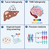 |
Figure 1 Multidimensional strategies for dissecting liver cancer heterogeneity and guiding precision medicine. (A) Tumor heterogeneity: Multi-region sampling and genomic profiling reveal branched evolutionary trajectories among intrahepatic lesions in primary liver cancer. (B) TIME heterogeneity: Single-cell and spatial omics technologies uncover diverse immune and stromal cell subsets shaping the TIME. (C) Organoid-based drug screening: Patient-derived organoids enable high-throughput evaluation of drug sensitivity and resistance mechanisms. (D) Precision medicine: Integrating tumor and TIME profiling with organoid-guided screening facilitates personalized treatment strategies for liver cancer patients. The image was created by Biorender. |
Tumor heterogeneity in primary liver cancer
Understanding the heterogeneity of liver cancer is crucial for elucidating tumor evolution and guiding therapeutic strategies [11, 12]. We have undertaken a series of studies to systematically dissect it at the tissue, single-cell, and phenotypic levels.
PLC patients exhibit high recurrence rates and poor prognosis, largely attributable to the frequent emergence of intrahepatic metastases. Nevertheless, the mechanisms driving intrahepatic metastasis and the clonal relationships among these distinct lesions have yet to be fully elucidated. We analyzed 43 tumor lesions from 10 hepatitis B virus (HBV)-related HCC patients by comparing the genomic information of different lesions within individual patients. We reconstructed an evolutionary tree illustrating the relationships among lesions, revealing a clear pattern of branched evolution among distinct lesions in liver cancer and supporting the notion that intrahepatic metastases often arise from a common ancestral clone [13]. Building on this, we systematically investigated the genomic features of liver cancer at the single-cell level. We proposed a dual-phase copy number evolution (DPCNE) model for the first time. Notably, we provided the single-cell evidence that polyploid liver cancers originate from diploid cells and identified CAD as a novel prognostic biomarker [14].
At the phenotypic level of tumor heterogeneity, cHCC-ICC contains both hepatocellular carcinoma and cholangiocarcinoma components and represents the subtype of liver cancer with the poorest prognosis. Currently, its molecular characteristics remain largely unknown, and accurate diagnostic biomarkers are lacking. Therefore, we profiled 133 cases of cHCC-ICC patients’ samples, identifying distinct molecular and clinical features across subtypes. Additionally, combined and mixed cHCC-ICCs exhibited monoclonal origins and high Nestin expression, suggesting potential diagnostic value [15]. What’s more, the latest study has shown that large duct (LD) and small duct (SD) subtypes of intrahepatic cholangiocarcinoma (iCCA) are characterized by distinct molecular alterations. LD-iCCA is commonly associated with mutations in KRAS and TP53, whereas SD-iCCA more frequently harbors IDH1/2 mutations and FGFR2 fusions [16].
The heterogeneity of tumor immune microenvironment in liver cancer
The clinical relevance of distinct TIME subtypes in liver cancer remains poorly understood, despite growing recognition of their potential impact on disease progression and therapeutic response [17]. Recently, we utilized single-cell RNA sequencing on 189 samples from 124 liver cancer patients and 8 mice, dissecting TIME heterogeneity [18]. Specifically, a total of 160 samples were collected from 124 liver cancer patients, including 10 blood samples, 14 adjacent tissues, 116 tumor tissues, and 20 metastatic samples. Additionally, 19 samples were obtained from 8 mice, comprising 9 tumor tissues, 4 blood samples, 4 adjacent liver tissues, and 2 bone marrow samples. Our study systematically characterized tumor-associated neutrophils (TANs), a key cellular subset in liver cancer, in terms of their heterogeneity and immunosuppressive functions. Neutrophils are highly fragile cells that typically survive no more than a week in peripheral circulation, and previous single-cell studies in liver cancer have largely overlooked this population [19, 20]. Leveraging a rapid processing workflow and antibody-free enrichment strategy, our study successfully captured over 30,000 neutrophils and identified 11 distinct TAN subpopulations, delineating their intratumoral differentiation trajectories. Furthermore, five TIME subtypes and their molecular features were defined in liver cancer. Our study highlighted the role of TANs, particularly in the myeloid-cell-enriched subtype, which is linked to poor prognosis due to their immunosuppressive activities. Evidence from mouse models suggested that targeting TANs could be an effective strategy for immunotherapy, as neutrophil depletion reduced tumor progression.
Recently, the functional states and composition of tumor-infiltrating T cells [21], B cells [22–24], NK cells [25], neutrophils [26], and myeloid cells [27] across pan-cancer cohorts have been characterized. Tumor-specific immune subsets, such as CXCL13+ T cells [21], FCRL4+ tumor-associated atypical B cells [22], ITGAX+ atypical B cells [23], SPP1+ macrophages [27], DNAJB1+ tumor-associated NK cells [25], HLA-DR+ neutrophil [26], and LAMP3⁺ dendritic cells [28], have been identified and linked to patient prognosis and immunotherapy response, providing critical insights into tumor ecosystem heterogeneity. With the accumulation of single-cell sequencing data, large-scale integration and tumor microenvironment subtyping are becoming feasible, offering new opportunities for precision immunotherapy. In addition, emerging technologies such as spatial omics, single-cell multi-omics, and CRISPR-based single-cell screens are further advancing our understanding of tumor microenvironment composition and function.
In addition to immune cell heterogeneity, vascular heterogeneity also plays a critical role in shaping the tumor microenvironment and treatment responses in liver cancer. Distinct vascular phenotypes, including capillarized, sinusoidal-like, and arterialized vasculature, are differentially associated with tumor subtypes and therapeutic vulnerabilities. For instance, tumors with arterialized vasculature may respond differently to anti-angiogenic therapies compared to those with sinusoidal remodeling [29]. Moreover, recent advances in imaging and histopathology have highlighted how the spatial and functional heterogeneity of liver tumor vasculature impacts drug delivery and immune infiltration [30, 31]. While this review focuses on immunologic aspects, the interplay between vascular architecture and immune contexture represents an important frontier in liver cancer research.
Applications of tumor organoids in precision medicine
Organoids faithfully retain the structural and genomic features of their tissues of origin and have become indispensable three-dimensional models in cancer research [32–37]. In recent years, compared to traditional two-dimensional cell lines and patient-derived xenograft (PDX) models, organoids have demonstrated unique advantages in drug screening and precision medicine. Notably, gene expression profiles at the transcriptomic level provide an important basis for drug sensitivity assessment using organoids [35, 36, 38].
We established a biobank of 399 tumor organoids from 144 patients to model the phenotypic and pharmacological heterogeneity of liver cancer. Through integrative analysis, we identified gene signatures predictive of treatment response and uncovered the role of c-Jun in lenvatinib resistance. Our study led to the development of PKUF-01, a novel compound showing synergistic effects in overcoming resistance [39]. Importantly, by comparing the drug responses of liver cancer organoids and corresponding patients undergoing standard clinical treatments, we found that the in vitro drug sensitivity of organoids closely mirrored the clinical responses observed in patients, highlighting the potential of organoid models to predict individual patient outcomes. Additionally, patient-derived organoids have been shown to faithfully recapitulate clinical drug responses, demonstrating high sensitivity, specificity, and predictive accuracy in forecasting patient outcomes to both targeted therapies and chemotherapeutic agents in breast and gastrointestinal cancer [36, 40].
Furthermore, organoids have become valuable tools for large-scale drug and toxicity screening. Integration with microfluidic systems, such as organoid-on-a-chip platforms, enables high-throughput drug testing while preserving key features of the tumor microenvironment [41]. In addition to identifying effective therapies, patient-derived organoids allow tracking of drug resistance evolution, as demonstrated by models replicating regorafenib resistance in colorectal cancer [40]. Moreover, organoids from adjacent normal tissues have been employed to predict drug-induced hepatotoxicity, nephrotoxicity, and cardiotoxicity, offering a comprehensive approach to optimizing both efficacy and safety in personalized treatment strategies [42–45].
Recent developments in immune-organoid co-culture systems have enabled more accurate modeling of immune responses. For example, Jacob et al. co-cultured glioblastoma organoids with CAR-T cells targeting EGFRvIII, demonstrating antigen-specific T cell activation and tumor cell killing [46]. Similarly, Neal et al. established immune-enhanced tumor organoids to evaluate the efficacy of the anti-PD-1 antibody nivolumab, showing induction of cytotoxic T cells, consistent with clinical responses. These studies underscore the potential of organoid platforms for rapid evaluation of immunotherapeutic strategies [47].
Discussion
Our recent studies have significantly deepened the understanding of genomic, phenotypic, and immunological heterogeneity underlying PLC. Through integrative approaches combining multi-region sequencing, single-cell technologies, and patient-derived organoid models, our studies have delineated the evolutionary dynamics of tumor lesions, uncovered novel prognostic biomarkers such as CAD. Also, we identified Nestin as the biomarker of cHCC-ICC. Concurrently, dissection of the TIME has revealed diverse and functionally pro-tumor neutrophils, highlighting their roles in shaping therapeutic outcomes. Notably, organoid platforms have emerged as powerful tools for modeling tumor diversity, predicting patient-specific drug responses, and enabling the functional evaluation of targeted and immunotherapies. Together, these findings provide a comprehensive framework for understanding the complexity of PLC and offer promising avenues for translating biological insights into personalized clinical strategies.
However, significant challenges remain in both the clinical and translational aspects of liver cancer research. Firstly, although organoids offer significant advantages over traditional cancer models and have developed rapidly, issues such as limited passaging capacity, low cryopreservation recovery rates, and restricted drug screening opportunities for patients persist. Challenges also exist in balancing fibroblast inclusion to better mimic the tumor microenvironment without allowing fibroblast overgrowth. Also, while liver cancer organoids have significantly advanced our understanding of tumor heterogeneity, drug response, and immune interactions, their ability to faithfully recapitulate metastatic behavior remains limited. Metastasis is a critical determinant of patient prognosis and the primary cause of liver cancer-related mortality, yet most organoid models are derived from primary tumor tissues and are cultured in isolation, lacking the cellular and structural components necessary for modeling the metastatic cascade. Recent efforts have attempted to address this limitation by deriving organoids from metastatic lesions, incorporating stromal or immune cell co-cultures, and applying organoid-on-a-chip or microfluidic platforms to mimic circulation and invasion dynamics. However, a robust and standardized organoid model capable of simulating the full metastatic potential of liver cancer cells is still lacking. Future work should prioritize the integration of spatial, mechanical, and immune cues into organoid systems, as well as the use of longitudinal patient sampling to trace clonal evolution during disease progression and metastasis. Secondly, the multi-omics datasets have only been partially analyzed. A large portion of the data remains unexplored and needs in-depth mining to uncover novel mechanisms of tumor immune regulation. Moreover, our study has identified several promising therapeutic targets, including PD-L1+TANs, the cHCC-ICC biomarker Nestin, and the molecular marker CAD linked to tumor heterogeneity. While these findings offer potential for therapeutic intervention, they still require validation in large-scale clinical cohorts. Further efforts are needed to develop diagnostic kits and conduct comprehensive translational studies, ultimately bridging basic research and clinical application to benefit patients.
Thus, our laboratory will focus on three key areas of research in the future. Firstly, we will investigate novel mechanisms by integrating single-cell multi-omics data with animal models and clinical data. This will enable a deeper understanding of the TIME, focusing on tumor-associated neutrophils [26, 48–50], fibroblasts [51–55], B cells [22, 23, 56–59], and NK cells [25, 60–62]. Secondly, we aim to identify new therapeutic targets and strategies. By mapping single-cell profiles across disease stages and treatment responses, and combining organoid models [63, 64] with CRISPR screening [65–67], we will validate candidate targets and evaluate them in representative mouse models. Lastly, we will enhance clinical translation by building a larger cancer cohort with major hospitals. Integrating clinical and preclinical data will support target validation and early clinical trials, aiming to improve clinical outcomes and advance precision oncology.
Funding
This project was supported by grants from the National Natural Science Foundation of China (No. 82030079, 81972656, 82341005, and 81988101 to N.Z.), the National Science and Technology Major Project of China (2022YFC3400900 to N.Z.), Baidu Foundation of China (No. 2020BD015 to N.Z.).
Conflicts of interest
The authors declare no conflicts of interest.
Data availability statement
No new data were generated or analyzed in this study.
Author contribution statement
Yinuo Wang and Liheng Chen contributed to the conceptualization and writing of the manuscript. Ning Zhang provided supervision and guidance throughout the writing process. All authors reviewed and approved the final version of the manuscript.
Ethics approval
This article is a review and does not contain any studies with human participants or animals performed by any of the authors. Therefore, ethics approval was not required.
References
- Sung H, Ferlay J, Siegel RL, Laversanne M, Soerjomataram I, Jemal A, et al. Global cancer statistics 2020: GLOBOCAN estimates of incidence and mortality worldwide for 36 cancers in 185 countries. CA Cancer J Clin. 2021;71(3):209–249. [CrossRef] [PubMed] [Google Scholar]
- Zhou J, Sun H, Wang Z, Cong W, Zeng M, Zhou W, et al. Guidelines for the diagnosis and treatment of primary liver cancer (2022 edition). Liver Cancer. 2023;12(5):405–444. [Google Scholar]
- Villanueva A. Hepatocellular carcinoma. N Engl J Med. 2019;380(15):1450–1462. [Google Scholar]
- Llovet JM, Kelley RK, Villanueva A, Singal AG, Pikarsky E, Roayaie S, et al. Hepatocellular carcinoma. Nat Rev Dis Primers. 2021;7(1):6. [Google Scholar]
- Dagogo-Jack I, Shaw AT. Tumour heterogeneity and resistance to cancer therapies. Nat Rev Clin Oncol. 2018;15(2):81–94. [Google Scholar]
- Chaisaingmongkol J, Budhu A, Dang H, Rabibhadana S, Pupacdi B, Kwon SM, et al. Common molecular subtypes among asian hepatocellular carcinoma and cholangiocarcinoma. Cancer Cell. 2017;32(1):57–70.e3. [Google Scholar]
- Dong L, Lu D, Chen R, Lin Y, Zhu H, Zhang Z, et al. Proteogenomic characterization identifies clinically relevant subgroups of intrahepatic cholangiocarcinoma. Cancer Cell. 2022;40(1):70–87.e15. [Google Scholar]
- Binnewies M, Roberts EW, Kersten K, Chan V, Fearon DF, Merad M, et al. Understanding the tumor immune microenvironment (TIME) for effective therapy. Nat Med. 2018;24(5):541–550. [Google Scholar]
- Li X, Ramadori P, Pfister D, Seehawer M, Zender L, Heikenwalder M. The immunological and metabolic landscape in primary and metastatic liver cancer. Nat Rev Cancer. 2021;21(9):541–557. [Google Scholar]
- Finn RS, Qin S, Ikeda M, Galle PR, Ducreux M, Kim TY, et al. Atezolizumab plus bevacizumab in unresectable hepatocellular carcinoma. N Engl J Med. 2020;382(20):1894–1905. [Google Scholar]
- Lee SH, Yim SY, Jeong YS, Li QX, Kang SH, Sohn BH, et al. Consensus subtypes of hepatocellular carcinoma associated with clinical outcomes and genomic phenotypes. Hepatology. 2022;76(6):1634–1648. [Google Scholar]
- Vitale I, Shema E, Loi S, Galluzzi L. Intratumoral heterogeneity in cancer progression and response to immunotherapy. Nat Med. 2021;27(2):212–224. [Google Scholar]
- Xue R, Li R, Guo H, Guo L, Su Z, Ni X, et al. Variable intra-tumor genomic heterogeneity of multiple lesions in patients with hepatocellular carcinoma. Gastroenterology. 2016;150(4):998–1008. [Google Scholar]
- Guo L, Yi X, Chen L, Zhang T, Guo H, Chen Z, et al. Single-cell DNA sequencing reveals punctuated and gradual clonal evolution in hepatocellular carcinoma. Gastroenterology. 2022;162(1):238–252. [Google Scholar]
- Xue R, Chen L, Zhang C, Fujita M, Li R, Yan SM, et al. Genomic and transcriptomic profiling of combined hepatocellular and intrahepatic cholangiocarcinoma reveals distinct molecular subtypes. Cancer Cell. 2019;35(6):932–947.e8. [Google Scholar]
- De Santis A, Zhu L, Tao J, Reißfelder C, Schölch S. Molecular subtypes of intrahepatic cholangiocarcinoma. Trends Mol Med. 2025;14:S1471-4914(25)00008-5. [Google Scholar]
- Thorsson V, Gibbs DL, Brown SD, Wolf D, Bortone DS, Ou Yang TH, et al. The immune landscape of cancer. Immunity. 2018;48(4):812–830.e14. [CrossRef] [PubMed] [Google Scholar]
- Xue R, Zhang Q, Cao Q, Kong R, Xiang X, Liu H, et al. Liver tumour immune microenvironment subtypes and neutrophil heterogeneity. Nature. 2022;612(7938):141–147. [Google Scholar]
- Shaul ME, Fridlender ZG. Tumour-associated neutrophils in patients with cancer. Nat Rev Clin Oncol. 2019;16(10):601–620. [Google Scholar]
- Ng LG, Ostuni R, Hidalgo A. Heterogeneity of neutrophils. Nat Rev Immunol. 2019;19(4):255–265. [Google Scholar]
- Zheng L, Qin S, Si W, Wang A, Xing B, Gao R, et al. Pan-cancer single-cell landscape of tumor-infiltrating T cells. Science. 2021;374(6574):abe6474. [Google Scholar]
- Yang Y, Chen X, Pan J, Ning H, Zhang Y, Bo Y, et al. Pan-cancer single-cell dissection reveals phenotypically distinct B cell subtypes. Cell. 2024;187(17):4790–4811.e22. [Google Scholar]
- Ma J, Wu Y, Ma L, Yang X, Zhang T, Song G, et al. A blueprint for tumor-infiltrating B cells across human cancers. Science. 2024;384(6695):eadj4857. [Google Scholar]
- Fitzsimons E, Qian D, Enica A, Thakkar K, Augustine M, Gamble S, et al. A pan-cancer single-cell RNA-seq atlas of intratumoral B cells. Cancer Cell. 2024;42(10):1784–1797.e4. [Google Scholar]
- Tang F, Li J, Qi L, Liu D, Bo Y, Qin S, et al. A pan-cancer single-cell panorama of human natural killer cells. Cell. 2023;186(19):4235–4251.e20. [Google Scholar]
- Wu Y, Ma J, Yang X, Nan F, Zhang T, Ji S, et al. Neutrophil profiling illuminates anti-tumor antigen-presenting potency. Cell. 2024;187(6):1422–1439.e24. [Google Scholar]
- Cheng S, Li Z, Gao R, Xing B, Gao Y, Yang Y, et al. A pan-cancer single-cell transcriptional atlas of tumor infiltrating myeloid cells. Cell. 2021;184(3):792–809.e23. [CrossRef] [PubMed] [Google Scholar]
- Zhang Q, He Y, Luo N, Patel SJ, Han Y, Gao R, et al. Landscape and dynamics of single immune cells in hepatocellular carcinoma. Cell. 2019;179(4):829–845.e20. [CrossRef] [PubMed] [Google Scholar]
- Fleischer JR, Jodszuweit CA, Ghadimi M, De Oliveira T, Conradi LC Vascular heterogeneity with a special focus on the hepatic microenvironment. Front Physiol. 2020;11:591901. [Google Scholar]
- Rhee H, Park YN, Choi JY. Advances in understanding hepatocellular carcinoma vasculature: implications for diagnosis, prognostication, and treatment. Korean J Radiol. 2024;25(10):887–901. [Google Scholar]
- Chen JA, Shi M, Li JQ, Qian CN. Angiogenesis: multiple masks in hepatocellular carcinoma and liver regeneration. Hepatol Int. 2010;4(3):537–547. [Google Scholar]
- Gao D, Vela I, Sboner A, Iaquinta PJ, Karthaus WR, Gopalan A, et al. Organoid cultures derived from patients with advanced prostate cancer. Cell. 2014;159(1):176–187. [CrossRef] [PubMed] [Google Scholar]
- Sato T, Vries RG, Snippert HJ, van de Wetering M, Barker N, Stange DE, et al. Single Lgr5 stem cells build crypt-villus structures in vitro without a mesenchymal niche. Nature. 2009;459(7244):262–265. [CrossRef] [PubMed] [Google Scholar]
- Barker N, Ridgway RA, van Es JH, van de Wetering M, Begthel H, van den Born M, et al. Crypt stem cells as the cells-of-origin of intestinal cancer. Nature. 2009;457(7229):608–611. [Google Scholar]
- van de Wetering M, Francies HE, Francis JM, Bounova G, Iorio F, Pronk A, et al. Prospective derivation of a living organoid biobank of colorectal cancer patients. Cell. 2015;161(4):933–945. [CrossRef] [PubMed] [Google Scholar]
- Sachs N, de Ligt J, Kopper O, Gogola E, Bounova G, Weeber F, et al. A living biobank of breast cancer organoids captures disease heterogeneity. Cell. 2018;172(1–2):373–386.e10. [CrossRef] [PubMed] [Google Scholar]
- Yan HHN, Siu HC, Law S, Ho SL, Yue SSK, Tsui WY, et al. A comprehensive human gastric cancer organoid biobank captures tumor subtype heterogeneity and enables therapeutic screening. Cell Stem Cell. 2018;23(6):882–897.e11. [CrossRef] [PubMed] [Google Scholar]
- Broutier L, Mastrogiovanni G, Verstegen MM, Francies HE, Gavarró LM, Bradshaw CR, et al. Human primary liver cancer-derived organoid cultures for disease modeling and drug screening. Nat Med. 2017;23(12):1424–1435. [CrossRef] [PubMed] [Google Scholar]
- Yang H, Cheng J, Zhuang H, Xu H, Wang Y, Zhang T, et al. Pharmacogenomic profiling of intra-tumor heterogeneity using a large organoid biobank of liver cancer. Cancer Cell. 2024;42(4):535–551,e8. [Google Scholar]
- Vlachogiannis G, Hedayat S, Vatsiou A, Jamin Y, Fernández-Mateos J, Khan K, et al. Patient-derived organoids model treatment response of metastatic gastrointestinal cancers. Science. 2018;359(6378):920–926. [CrossRef] [PubMed] [Google Scholar]
- Skardal A, Shupe T, Atala A. Organoid-on-a-chip and body-on-a-chip systems for drug screening and disease modeling. Drug Discov Today. 2016;21(9):1399–1411. [Google Scholar]
- Drost J, Clevers H. Organoids in cancer research. Nat Rev Cancer. 2018;18(7):407–418. [Google Scholar]
- Morizane R, Lam AQ, Freedman BS, Kishi S, Valerius MT, Bonventre JV. Nephron organoids derived from human pluripotent stem cells model kidney development and injury. Nat Biotechnol. 2015;33(11):1193–1200. [Google Scholar]
- Eder A, Vollert I, Hansen A, Eschenhagen T. Human engineered heart tissue as a model system for drug testing. Adv Drug Deliv Rev. 2016;96:214–224. [Google Scholar]
- Voges HK, Mills RJ, Elliott DA, Parton RG, Porrello ER, Hudson JE. Development of a human cardiac organoid injury model reveals innate regenerative potential. Development. 2017;144(6):1118–1127. [Google Scholar]
- Jacob F, Salinas RD, Zhang DY, Nguyen PTT, Schnoll JG, Wong SZH, et al. A patient-derived glioblastoma organoid model and biobank recapitulates inter- and intra-tumoral heterogeneity. Cell. 2020;180(1):188–204.e22. [Google Scholar]
- Neal JT, Li X, Zhu J, Giangarra V, Grzeskowiak CL, Ju J, et al. Organoid modeling of the tumor immune microenvironment. Cell. 2018;175(7):1972–1988.e16. [CrossRef] [PubMed] [Google Scholar]
- Gungabeesoon J, Gort-Freitas NA, Kiss M, Bolli E, Messemaker M, Siwicki M, et al. A neutrophil response linked to tumor control in immunotherapy. Cell. 2023;186(7):1448–1464.e20. [Google Scholar]
- Ng MSF, Kwok I, Tan L, Shi C, Cerezo-Wallis D, Tan Y, et al. Deterministic reprogramming of neutrophils within tumors. Science. 2024;383(6679):eadf6493. [Google Scholar]
- Benguigui M, Cooper TJ, Kalkar P, Schif-Zuck S, Halaban R, Bacchiocchi A, et al. Interferon-stimulated neutrophils as a predictor of immunotherapy response. Cancer Cell. 2024;42(2):253–265.e12. [Google Scholar]
- Krishnamurty AT, Shyer JA, Thai M, Gandham V, Buechler MB, Yang YA, et al. LRRC15(+) myofibroblasts dictate the stromal setpoint to suppress tumour immunity. Nature. 2022;611(7934):148–154. [Google Scholar]
- Hutton C, Heider F, Blanco-Gomez A, Banyard A, Kononov A, Zhang X, et al. Single-cell analysis defines a pancreatic fibroblast lineage that supports anti-tumor immunity. Cancer Cell. 2021;39(9):1227–1244.e20. [Google Scholar]
- Gao Y, Li J, Cheng W, Diao T, Liu H, Bo Y, et al. Cross-tissue human fibroblast atlas reveals myofibroblast subtypes with distinct roles in immune modulation. Cancer Cell. 2024;42(10):1764–1783.e10. [Google Scholar]
- Liu Y, Sinjab A, Min J, Han G, Paradiso F, Zhang Y, et al. Conserved spatial subtypes and cellular neighborhoods of cancer-associated fibroblasts revealed by single-cell spatial multi-omics. Cancer Cell. 2025;43(5):905–924.e6. [Google Scholar]
- Cords L, de Souza N, Bodenmiller B. Classifying cancer-associated fibroblasts – the good, the bad, and the target. Cancer Cell. 2024;42(9):1480–1485. [CrossRef] [PubMed] [Google Scholar]
- Cabrita R, Lauss M, Sanna A, Donia M, Skaarup Larsen M, Mitra S, et al. Tertiary lymphoid structures improve immunotherapy and survival in melanoma. Nature. 2020;577(7791):561–565. [Google Scholar]
- Helmink BA, Reddy SM, Gao J, Zhang S, Basar R, Thakur R, et al. B cells and tertiary lymphoid structures promote immunotherapy response. Nature. 2020;577(7791):549–555. [Google Scholar]
- Petitprez F, de Reynies A, Keung EZ, Chen TW, Sun CM, Calderaro J, et al. B cells are associated with survival and immunotherapy response in sarcoma. Nature. 2020;577(7791):556–560. [CrossRef] [PubMed] [Google Scholar]
- Fridman WH, Meylan M, Pupier G, Calvez A, Hernandez I, Sautes-Fridman C. Tertiary lymphoid structures and B cells: An intratumoral immunity cycle. Immunity. 2023;56(10):2254–2269. [Google Scholar]
- Yan J, Zhang C, Xu Y, Huang Z, Ye Q, Qian X, et al. GPR34 is a metabolic immune checkpoint for ILC1-mediated antitumor immunity. Nat Immunol. 2024;25(11):2057–2067. [Google Scholar]
- Delconte RB, Owyong M, Santosa EK, Srpan K, Sheppard S, McGuire TJ, et al. Fasting reshapes tissue-specific niches to improve NK cell-mediated anti-tumor immunity. Immunity. 2024;57(8):1923–1938.e7. [Google Scholar]
- Tarannum M, Ding X, Barisa M, Hu S, Anderson J, Romee R, et al. Engineering innate immune cells for cancer immunotherapy. Nat Biotechnol. 2025;43(4):516–533. [Google Scholar]
- Zhou Z, Van der Jeught K, Fang Y, Yu T, Li Y, Ao Z, et al. An organoid-based screen for epigenetic inhibitors that stimulate antigen presentation and potentiate T-cell-mediated cytotoxicity. Nat Biomed Eng. 2021;5(11):1320–1335. [Google Scholar]
- Du Y, Wang YR, Bao QY, Xu XX, Xu C, Wang S, et al. Personalized vascularized tumor organoid-on-a-chip for tumor metastasis and therapeutic targeting assessment. Adv Mater. 2025;37(6):e2412815. [Google Scholar]
- Luo C, Zhang R, Guo R, Wu L, Xue T, He Y, et al. Integrated computational analysis identifies therapeutic targets with dual action in cancer cells and T cells. Immunity. 2025;58(3):745–765.e9. [Google Scholar]
- Zhou P, Shi H, Huang H, Sun X, Yuan S, Chapman NM, et al. Single-cell CRISPR screens in vivo map T cell fate regulomes in cancer. Nature. 2023;624(7990):154–163. [Google Scholar]
- Moffat J, Komor AC, Lum L. Impact of CRISPR in cancer drug discovery. Science. 2024;386(6720):378–379. [Google Scholar]
Cite this article as: Wang Y, Chen L & Zhang N. Advances in basic and translational research on the heterogeneity and metastatic mechanisms of primary liver cancer. Visualized Cancer Medicine. 2025; 6, 10. https://doi.org/10.1051/vcm/2025012.
All Figures
|
Figure 1 Multidimensional strategies for dissecting liver cancer heterogeneity and guiding precision medicine. (A) Tumor heterogeneity: Multi-region sampling and genomic profiling reveal branched evolutionary trajectories among intrahepatic lesions in primary liver cancer. (B) TIME heterogeneity: Single-cell and spatial omics technologies uncover diverse immune and stromal cell subsets shaping the TIME. (C) Organoid-based drug screening: Patient-derived organoids enable high-throughput evaluation of drug sensitivity and resistance mechanisms. (D) Precision medicine: Integrating tumor and TIME profiling with organoid-guided screening facilitates personalized treatment strategies for liver cancer patients. The image was created by Biorender. |
| In the text | |
Current usage metrics show cumulative count of Article Views (full-text article views including HTML views, PDF and ePub downloads, according to the available data) and Abstracts Views on Vision4Press platform.
Data correspond to usage on the plateform after 2015. The current usage metrics is available 48-96 hours after online publication and is updated daily on week days.
Initial download of the metrics may take a while.